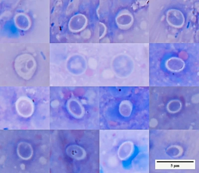
If fish are dying in the aquarium for unknown reasons:
- Transfer all fish to another tank.
- Investigate what is happening.
Mass mortalities can vary:
Different species are dying simultaneously.
Different genera and families have distinct lifestyles, physiology, and immune responses. If multiple species die at once, the cause is more likely water chemistry rather than a pathogen.
One species dies quickly and massively.
This is also often related to water chemistry — the most sensitive species reacts first.
One species dies periodically, over time.
In this case, infections or parasites are more likely, especially if this species has lower resistance (e.g., ciliates, bacteria).
If several fish have died in the same tank (a mass mortality event):
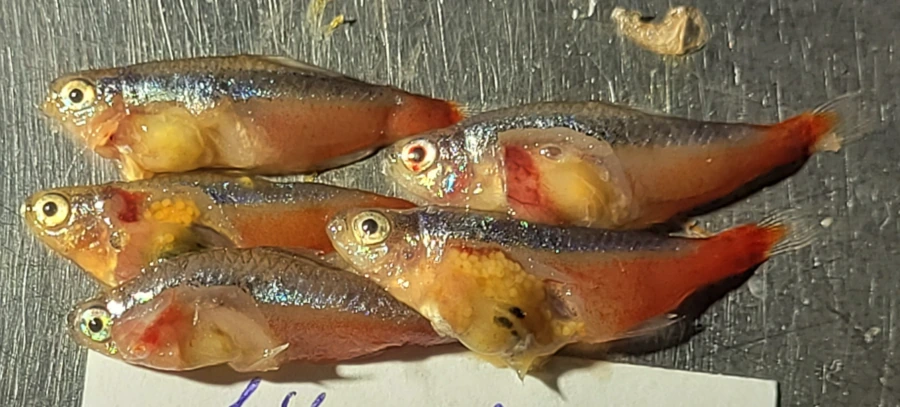
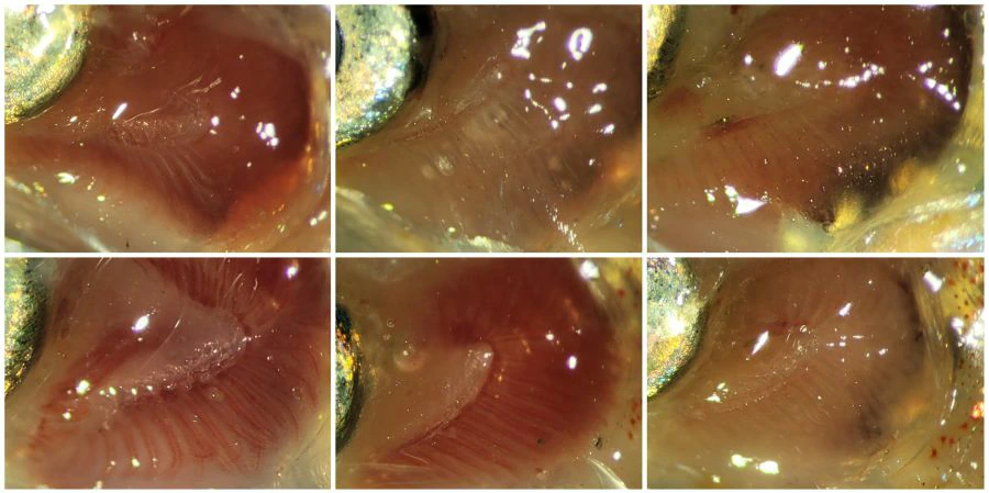
Examine several fish at once for comparison and to distinguish common changes from individual pathology. During a mass mortality event, dead fish should ideally be grouped by the time they were found, and that time should be recorded. This makes it possible to compare early and late cases and reconstruct the sequence of events more accurately.
View the neon tetra liver slide in the virtual microscope
A fish’s appetite is not an indicator of health
Fish have very strong instincts. Even if a fish eats, it does not mean it is healthy.
The intestine may be non-functional (smooth, transparent, like a water-filled bag).
Such a fish eats but dies of starvation: the stomach and intestine contain no food fragments.
It is possible the fish vomits after feeding — but this often goes unnoticed.
Do not rely on appetite as a criterion of health.
If transferring fish from “dirty” water to clean water:
Treat them for bacterial infections. Otherwise, the disease is likely to manifest later.
If the fish has been exposed to Cryptocarion irritans:
Take regular scrapes and washes from the gills (at least once a month).
The gill form is invisible on the body… until it is too late.Cryptocaryon irritans: Not Just White Spots Cryptocaryon irritans. is a ciliated protozoan parasite, often visible to the naked eye. Tangs, butterflyfish, and boxfish are particularly …
Fish can be handled and treated directly
Fish should not be injured unnecessarily. However, this does not mean that they cannot be restrained, examined, or treated by hand.
Brief restraint with wet hands in examination gloves is a routine practical procedure. It allows the operator to collect a skin scrape, treat a wound, remove necrotic tissue, fungal growth, external parasites, or excessive pathologically altered mucus.
If a parasite or affected material is located on the body surface and is mechanically accessible, direct removal should at least be considered. Even incomplete cleaning can substantially reduce the burden on the fish within a few minutes.
Infected mucus, deteriorating epithelium, or necrotic tissue should not be preserved merely because the mucus layer normally performs a protective function. In a particular case, this material may already contain large numbers of parasites, bacteria, and tissue debris.
The restriction should apply not to touching the fish itself, but to handling it incorrectly.
If an external parasite can be removed, remove it
There is no need to wait for a treatment dissolved in the water to affect the entire parasite burden.
Ciliates, external worms, parasitic crustaceans, fungal hyphae growing on dead tissue, and other accessible objects can be removed by rinsing, gentle wiping, forceps, or another suitable method. The choice of technique depends on the fish species, the structure of its body surface, the location of the lesion, and the condition of the animal.
Practical example: mechanical reduction of the parasite burden in cryptocaryonosisThe inability to remove every parasite in a single procedure is not a reason to leave all parasites on the fish.
 Cryptocaryonosis: killing the parasite or treating the water?
A practical analysis of cryptocaryonosis treatment in marine fish: why killing the parasite should not become more important than the …
Cryptocaryonosis: killing the parasite or treating the water?
A practical analysis of cryptocaryonosis treatment in marine fish: why killing the parasite should not become more important than the …
If a fish dies during non-traumatic handling (bathing, transfer):
Necropsy may reveal irreversible changes that could have caused death on their own.
Stress only triggered the death, which would have occurred soon anyway.
So don’t blame yourself immediately — perform a necropsy and then draw conclusions.
Most likely, the fish was already “doomed.”
If fungal hyphae (Saprolegnia) appear on the body :
More often, the oomycete attaches to necrotic areas of skin or gills.
If the hyphae look like white fluff, it is most likely a saprophyte — not the cause, but a consequence, an indicator.
In “aggressive” oomycetes, the hyphae spread along the skin. The coating is denser, resembling wet cotton, and individual filaments are not visible.
If fungal hyphae are present on the fish:
Treat bacterial infection at the same time.
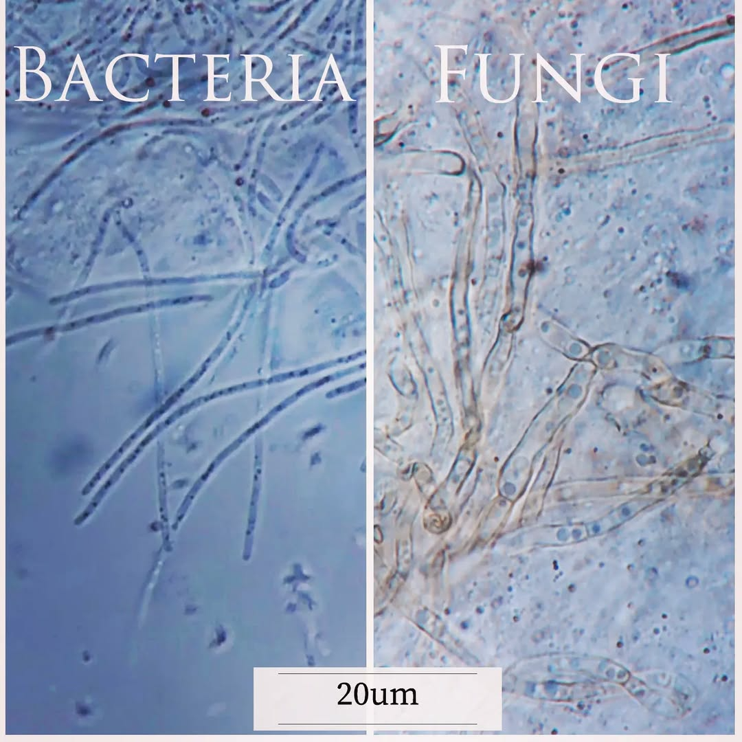
Bacteria, ciliates, and flagellates always thrive in these fungal “thickets.”
Do not confuse bacteria with fungi — the size difference is enormous.
FAQ
If a fish eats, is it healthy?
No. Appetite does not guarantee health.
Why do different species die at once?
Most often due to water conditions, not infection.
Should treatment start immediately after deaths?
No. The cause must be identified first.
How many fish should be examined?
Several, to distinguish individual from systemic issues.
Is Saprolegnia the cause of death?
Usually not. It is a secondary colonizer.
See also:
Gas bubble disease. Part I
 Gas Bubble Disease: It's Not About the Bubbles
Gas Bubble Disease in fish is not caused by air bubbles entering the gills but by dissolved gas supersaturation. This article explains the …
Gas Bubble Disease: It's Not About the Bubbles
Gas Bubble Disease in fish is not caused by air bubbles entering the gills but by dissolved gas supersaturation. This article explains the …
Granulomas and intracellular agents in fish
 Granulomas and intracellular agents in fish
A collection of real observations: intracellular yeast-like organisms, coccidia, cryptosporidia, and fungal structures in fish.
Granulomas and intracellular agents in fish
A collection of real observations: intracellular yeast-like organisms, coccidia, cryptosporidia, and fungal structures in fish.
Hexamita
 Hexamitids Without Myths
Why hexamitids are not a normal component of fish intestinal microflora, where the myth of 'internal parasites' comes from, and how …
Hexamitids Without Myths
Why hexamitids are not a normal component of fish intestinal microflora, where the myth of 'internal parasites' comes from, and how …
What to do with fish after arrival
 Initial Diagnostics After Fish Transportation
Microscopy, smears, staining methods, and common mistakes in early diagnostics.
Initial Diagnostics After Fish Transportation
Microscopy, smears, staining methods, and common mistakes in early diagnostics.