The examined fish showed no visible signs of disease during life. Mortality occurred predominantly at night.
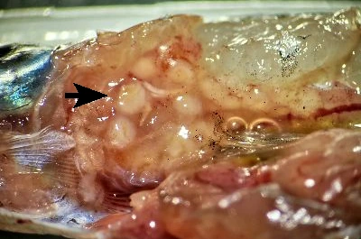
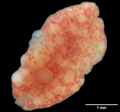
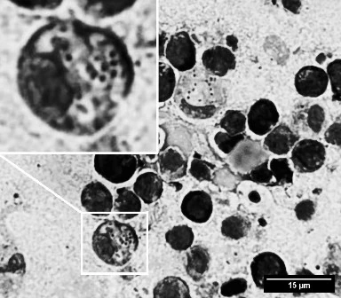
Gills were pale, and the fish were slightly emaciated. Necropsy revealed granulomas in the spleen and/or kidneys. Parenchymal organs were enlarged and anemic. The gross appearance was consistent with mycobacteriosis. However, Ziehl–Neelsen staining of impression smears from spleen and kidney did not reveal acid-fast bacteria. The granulomas were necrotic, a feature commonly associated with mycobacterial infections.
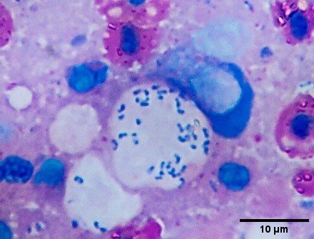
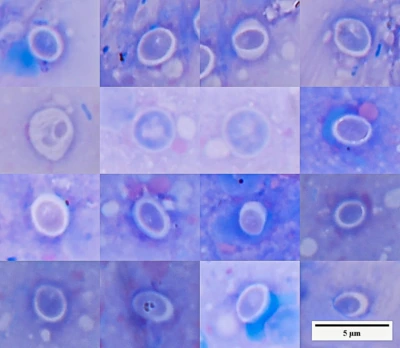
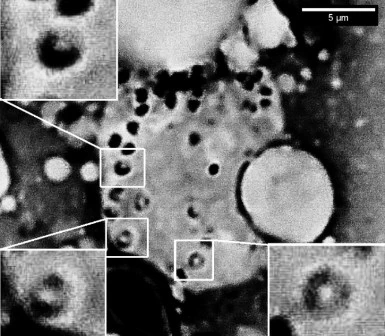
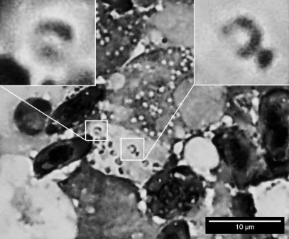
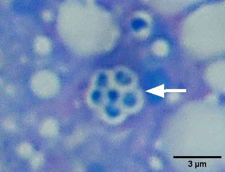
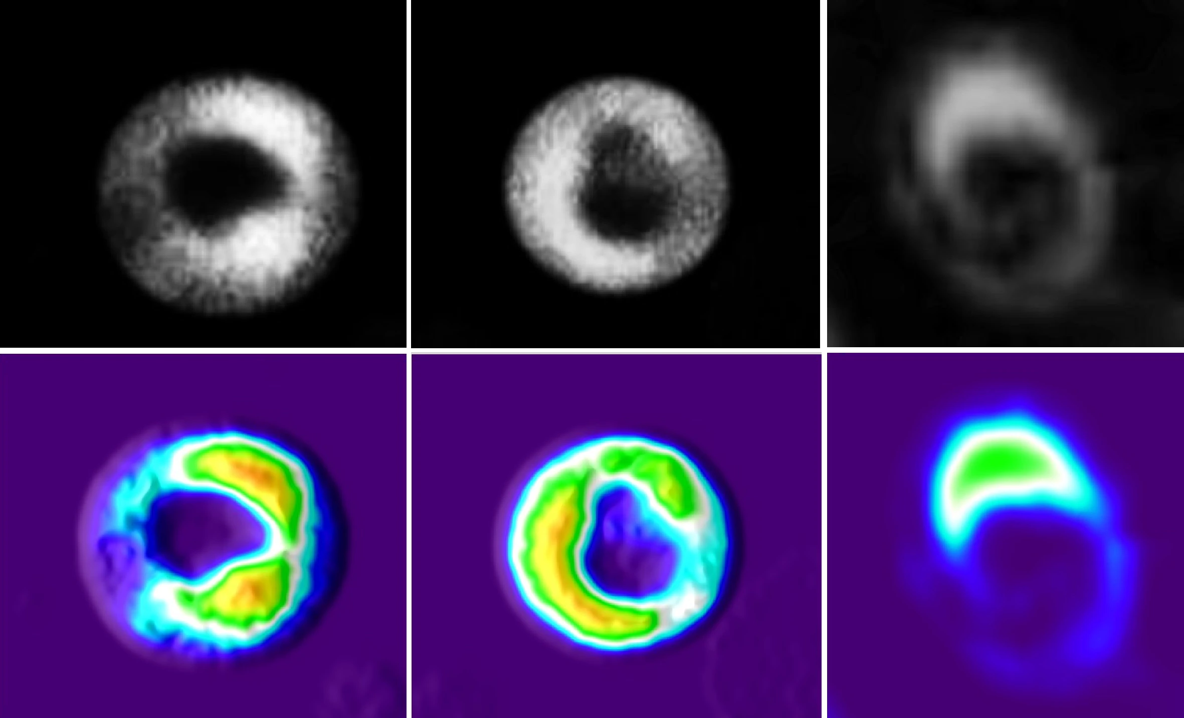
Staining of impression smears using Gram and Romanowsky–Giemsa methods, combined with careful microscopy, revealed numerous small Gram-positive budding cells (1–3 µm) within histiocytes of the kidney and spleen. Larger cells showed structures resembling basophilic, horseshoe-shaped nuclei (Fig. 1–6).

The findings are consistent with a diffuse invasive mycosis caused by yeast-like organisms resembling Histoplasma. The formation of epithelioid-cell granulomas and necrotizing granulomatous inflammation is commonly described in H. capsulatum infections. No mycelial forms were observed in any of the samples. The extent of granulomatous inflammation in parenchymal organs suggests a chronic, slowly progressing disease.
This case highlights the presence of diseases with atypical etiology in aquarium systems and emphasizes the importance of thorough pathogen identification. Available literature mainly describes experimental infections of poikilothermic animals aimed at studying histoplasmosis under temperatures lower than typical for this pathogen (below 35°C)(“Experimental histoplasmosis in cold-blooded animals” DOI:10.1007/BF02063081).