“Granulomas Found—So It Must Be Mycobacteriosis”
During fish necropsies, whitish nodules in the spleen, liver, or kidneys often raise suspicion of mycobacteriosis. This suspicion is reasonable: granulomatous lesions are indeed characteristic of many cases of mycobacterial infection. The mistake comes at the next step—when a morphological finding is treated as an already established etiology.
Three concepts need to be kept separate: a visible nodule, a granuloma, and mycobacteriosis. They are related, but they are not equivalent.
What Are We Actually Seeing?
Two photographs of fish spleens show numerous whitish lesions. Their gross appearances are similar, but microscopy reveals different findings.
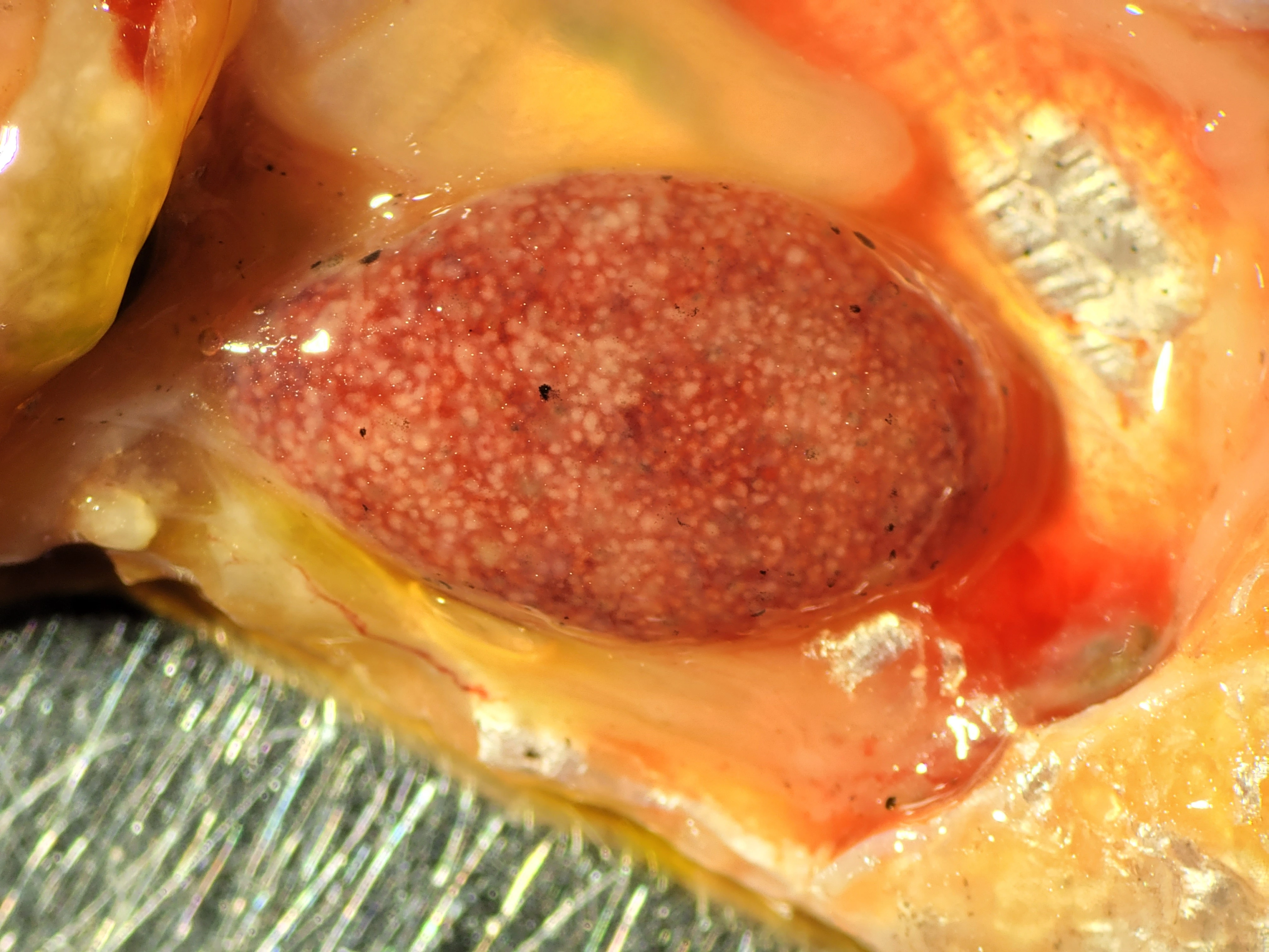
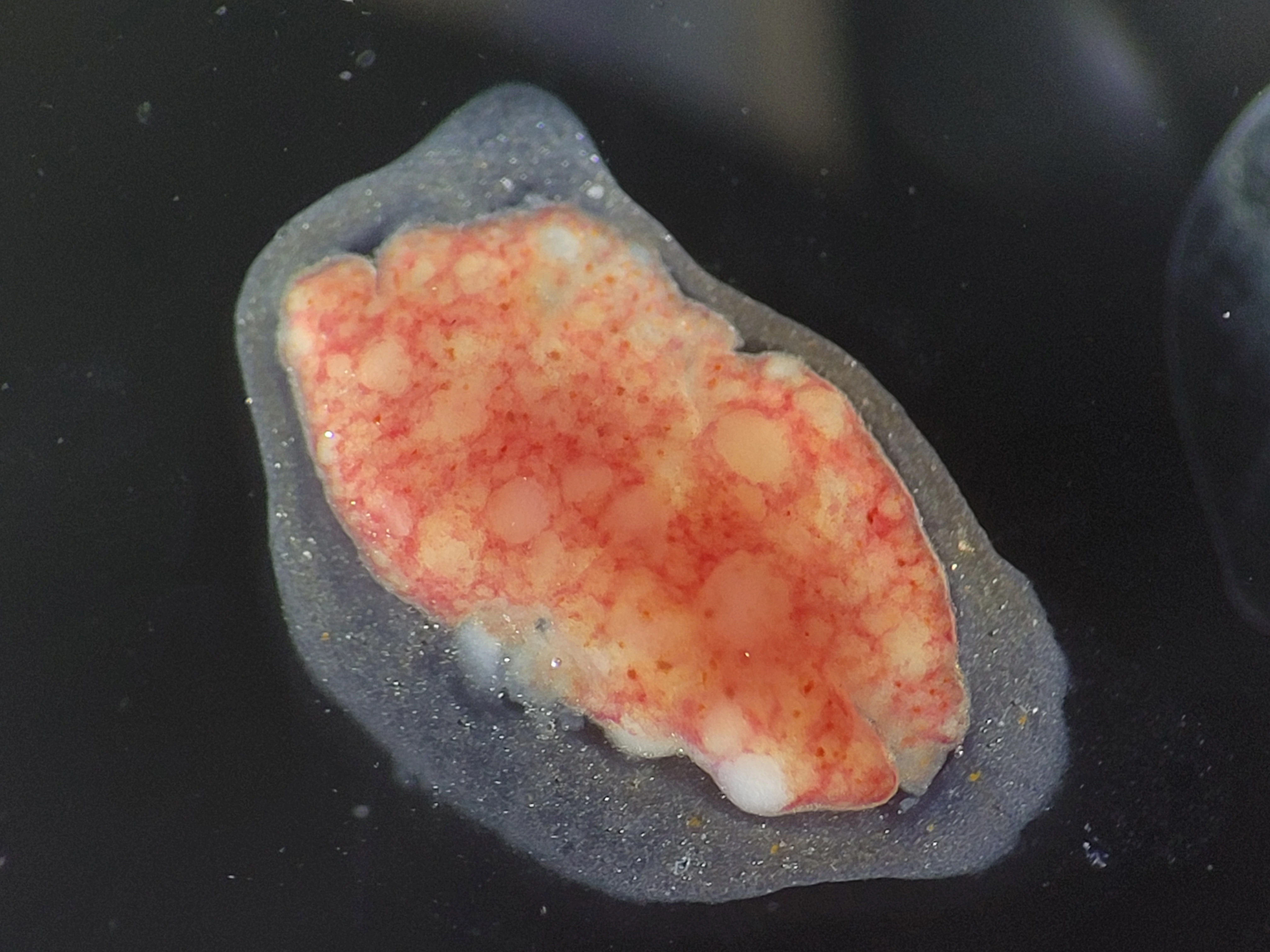
The color and distribution of visible lesions do not reveal their cellular architecture. Therefore, a white nodule seen at necropsy should not automatically be called a granuloma. The tissue must first be examined—for example, by histology.
What Is a Granuloma Made Of, and Why Does It Form?
In its classical definition, a granuloma is an organized collection of mature mononuclear phagocytes, primarily macrophages. These cells engulf and process microorganisms, foreign particles, and debris from damaged tissues. Other cells may be present in a granuloma, and its center may contain necrosis, but neither feature is required by the definition (Pagán & Ramakrishnan, 2018).
If a macrophage can eliminate an irritant, a prolonged organized reaction may not develop. But when material persists, is difficult to break down, or is too large, additional cells are recruited and a localized lesion forms.
Importantly, the persistent material does not necessarily come from outside the body. The reaction may also develop around remnants of the organism’s own damaged tissues. Thus, the equation “granuloma = infection” is incorrect even at the level of mechanism.
Granulomas can develop in response to various bacterial, fungal, and parasitic infections, as well as certain noninfectious processes. A granulomatous response describes the host’s reaction; its etiology must be established separately.
Why Shape and Necrosis Do Not Provide a Diagnosis
When examining granulomatous lesions, we assess their organization, size, number, the presence of necrosis, the prominence of peripheral cellular layers, and the development of a connective-tissue capsule.
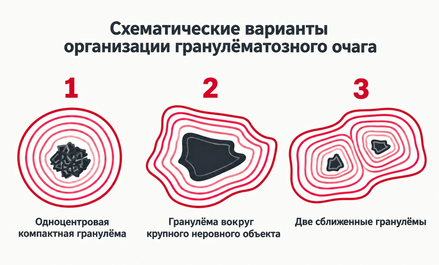
The full article presents a useful diagram with a single compact center, a large irregular center, and adjacent lesions that may merge. This is a model of possible patterns of organization, not a way to identify a pathogen reliably from a lesion’s outline.
Necrosis—the death of cells within a lesion—does occur in mycobacteriosis, but it is not unique to it. Conversely, the absence of necrosis does not rule out mycobacterial infection. The same applies to capsule thickness and the regularity of lesion contours.
Within a group of fish, similar lesions may develop a similar structure. This can help compare cases and suggest a shared pathological process, but it does not make any particular granuloma shape unique to a specific pathogen.
Not Every Round Structure Is a Granuloma
The full article shows structures in neon tetra kidneys that are tentatively interpreted as mineral concretions; some are surrounded by features resembling a granulomatous response. Fibrous nodular structures in gudgeon kidneys are also shown. These findings are deliberately described with caution: without further confirmation, not every nodule or granuloma-like cellular arrangement should be assigned to a specific pathological process.
View these specimens in the interactive microscope in the F.O.S. article.
Many Granulomas—Does That Mean a High Bacterial Load?
Not necessarily. The number and integrity of granulomas depend not only on the pathogen, but also on how the immune response is organized. Well-formed lesions may help contain infection. When this organization breaks down, mycobacteria may multiply extensively while the granulomas deteriorate.
Moreover, experimental models of Mycobacterium marinum infection in zebrafish have shown that, under certain conditions, mycobacteria can exploit the recruitment and movement of macrophages to spread further (Pagán & Ramakrishnan, 2015).
A granuloma, then, is not always a static capsule surrounding a pathogen, nor is it a “counter” of bacterial cells.
From a Finding to an Etiological Diagnosis
It helps to distinguish different levels of evidence.
- Gross examination. Whitish nodules or lesions are visible in particular organs. This describes their external appearance, not their cellular composition.
- Tissue microscopy. We assess whether granulomatous architecture is confirmed, what occupies the center of a lesion, and whether necrosis or other changes are present.
- Special staining. Ziehl–Neelsen staining can reveal structures that retain the dye after acid decolorization. Finding such structures, especially within lesions, strengthens the suspicion of mycobacteriosis. However, acid-fastness does not identify a bacterial species, and a negative stain alone does not rule out infection.
- Pathogen identification. When necessary, culture, PCR, sequencing, or a combination of methods may be used, taking sample quality and the clinical context into account.
For more on what this staining method actually tests, see “Tradition Instead of Science I. Method”.
This sequence does not imply that every test is available in every case. Its purpose is to make clear the level of certainty reached by our conclusion.
Where Is the Boundary?
“Multiple whitish lesions were found in the organs” is a gross observation.
“Granulomas were confirmed in the tissue” is a morphological conclusion.
“Acid-fast rod-shaped bacteria were detected” is the result of a special investigation.
“Mycobacteriosis was diagnosed” is an etiological diagnosis that requires consideration of the evidence as a whole.
We should move from one statement to the next as evidence becomes available—not merely because these findings often occur together.
A granuloma helps characterize a pathological process. Its appearance alone, however, cannot establish the cause.
References
- Pagán, A. J., Ramakrishnan, L. (2018). The Formation and Function of Granulomas. Annual Review of Immunology, 36, 639–665. https://doi.org/10.1146/annurev-immunol-032712-100022
- Pagán, A. J., Ramakrishnan, L. (2015). Immunity and Immunopathology in the Tuberculous Granuloma. Cold Spring Harbor Perspectives in Medicine, 5, a018499. https://doi.org/10.1101/cshperspect.a018499
Other versions: F. — the story of two spleens, without technical terminology · F.O.S. — the full analysis, examples, and interactive microscopy collections.
See also
colonization or invasive process
 Fungal structures in fish: colonization or invasive processFungal structures in fish may represent either surface colonization or an invasive process. This article discusses morphological features, …
Hexamitids Without Myths
Fungal structures in fish: colonization or invasive processFungal structures in fish may represent either surface colonization or an invasive process. This article discusses morphological features, …
Hexamitids Without Myths
 Hexamitids Without MythsWhy hexamitids are not a normal component of fish intestinal microflora, where the myth of 'internal parasites' comes from, and how …
Cryptocaryon irritans: Not Just
Hexamitids Without MythsWhy hexamitids are not a normal component of fish intestinal microflora, where the myth of 'internal parasites' comes from, and how …
Cryptocaryon irritans: Not JustWhite Spots
 Cryptocaryon irritans: Not Just White SpotsCryptocaryon irritans. is a ciliated protozoan parasite, often visible to the naked eye. Tangs, butterflyfish, and boxfish are particularly …
Health Does Not Mean
Cryptocaryon irritans: Not Just White SpotsCryptocaryon irritans. is a ciliated protozoan parasite, often visible to the naked eye. Tangs, butterflyfish, and boxfish are particularly …
Health Does Not MeanSterility
 Health Does Not Mean SterilityWhy the presence of microorganisms and parasites does not always mean disease, why immunity does not have to eliminate them completely, and …
Ichthyophthiriosis Without Myths
Health Does Not Mean SterilityWhy the presence of microorganisms and parasites does not always mean disease, why immunity does not have to eliminate them completely, and …
Ichthyophthiriosis Without Myths
 Ichthyophthiriosis Without MythsA science-based analysis of Ichthyophthirius multifiliis: life cycle, cyst terminology, the myth of parasite ubiquity, and real mechanisms …
Two Sides of One
Ichthyophthiriosis Without MythsA science-based analysis of Ichthyophthirius multifiliis: life cycle, cyst terminology, the myth of parasite ubiquity, and real mechanisms …
Two Sides of OneDosage
 Two Sides of One DosageAquarium medications: remedy or threat? Pros and cons of antibiotics, formalin, methylene blue, malachite green, and the FMC mixture.