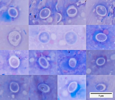
At least two clinical situations are possible in cryptocaryonosis.
In the first, the parasite is detected early: its numbers are still low, and the fish retains normal respiration, behaviour, and appetite. In this situation there is time to target the free-living stages in the water, control the concentration of the selected treatment, and gradually reduce contamination of the system.
In the second situation, the disease is already severe. The fish is breathing heavily, is covered with large amounts of mucus and parasites, refuses food, and may die before treatment of the water produces a result.
These situations are not opposed to one another. Successful assistance to a severely affected fish should move it from the second state into the first: first reduce the parasite burden rapidly and restore vital functions, then continue treatment of the system and control the remaining stages of the parasite.
This page is devoted specifically to the second situation — when the fish is already in distress.
Why the presence of a parasite does not always mean disease, and why health does not mean sterilityHealth Does Not Mean Sterility Why the presence of microorganisms and parasites does not always mean disease, why immunity does not have to eliminate them completely, and …
What should be done if the fish may die before the treatment in the water takes effect?
Adding copper to the water does not immediately remove trophonts from the skin and gills. Even a correctly calculated and controlled concentration acts within the parasite’s life cycle and requires time (Zec et al., 2021).
For the system, this may be a justified treatment.
For a fish that is suffocating, it is too slow as the first intervention.
This is not an argument against copper or other methods of treating the water. It is an argument about the sequence of actions. When the fish is severely affected, waiting for a drug to act should not replace direct assistance to the animal.
Treatment should therefore be divided into two stages:
- Immediate assistance to the fish and rapid reduction of the parasite burden;
- Subsequent control of the parasite in the water and prevention of reinfection.
Reduce the burden on the fish first
The severity of the condition is determined not only by the fact of infection, but also by the number of parasites present on the skin and gills at that moment. The greater the burden, the more severe the irritation of the body surface, mucus production, epithelial damage, and respiratory impairment (Kaige & Miyazaki, 1985; Cervera et al., 2022).
Therefore, when the condition is severe, the first objective is not to destroy every parasite in the system immediately, but to reduce their number directly on the fish as quickly as possible.
Even incomplete parasite removal can change the situation substantially. If only several hundred parasites remain on the skin and gills instead of thousands, the immune system, body surface, and respiratory organs gain time to recover. The fish may begin to breathe more effectively, maintain equilibrium, and survive the period required for further treatment.
Complete elimination of the parasite may take time.
Reduction of the burden on the fish should begin immediately.
Methods acting directly on the body surface may include:
- A short bath in an appropriate solution;
- Rinsing;
- Removal of excessive pathologically altered mucus;
- Careful mechanical cleaning of accessible areas of skin;
- Treatment of individual lesions;
- Transfer of the fish to a clean container after the procedure.
The method must be selected according to the fish species, the condition of its body surface, the location of the lesions, and the animal’s ability to tolerate restraint and treatment.
This does not mean roughly scraping normal epithelium. The material removed should primarily be material that has already ceased to perform a protective function: accumulations of parasites, loose mucus, detached epithelium, and superficial pathological deposits.
More information: direct handling of fish and mechanical removal of external parasites Simple Rules for Working with Aquarium Fish
Basic guidelines for diagnosing and caring for aquarium fish: what to do in case of mortality, how to interpret appetite, and when to treat …
Simple Rules for Working with Aquarium Fish
Basic guidelines for diagnosing and caring for aquarium fish: what to do in case of mortality, how to interpret appetite, and when to treat …
Why parasites appear on the fish in waves
Trophonts of Cryptocaryon irritans feed and grow on the skin and gills of the fish. After completing this stage, they leave the host, settle on surfaces, and continue their development outside the fish (Colorni & Diamant, 1993; Ma et al., 2016).
In an aquarium, these stages do not disappear. They accumulate on the bottom, walls, equipment, and other surfaces. After division, new infective stages emerge and search for a host. Recently emerged theronts pose the greatest risk: as they age, their motility and ability to infect fish decline rapidly (Yoshinaga & Dickerson, 1994; Chi et al., 2020).
The number of visible parasites on a fish may therefore change in waves:
- Some trophonts leave the fish, and its external appearance temporarily improves;
- Development continues outside the host;
- A new generation emerges from the accumulated stages;
- The fish becomes reinfected, sometimes much more heavily than before.
A temporary decrease in the number of white spots on the fish does not necessarily mean that the disease has ended.
It may simply reflect the transition of the parasite to the next stage of its life cycle (Li et al., 2022).
The fish remains close to the site of parasite development
After leaving the host, some stages of Cryptocaryon irritans settle, attach to aquarium surfaces, and continue developing outside the fish (Colorni, 1985; Colorni & Diamant, 1993). Fish may remain directly on or close to the bottom for long periods, only a few centimetres from accumulations of the parasite. The host therefore remains close to the site from which the next generation of infective stages emerges. The limited water volume and short distance increase the probability of reinfection.
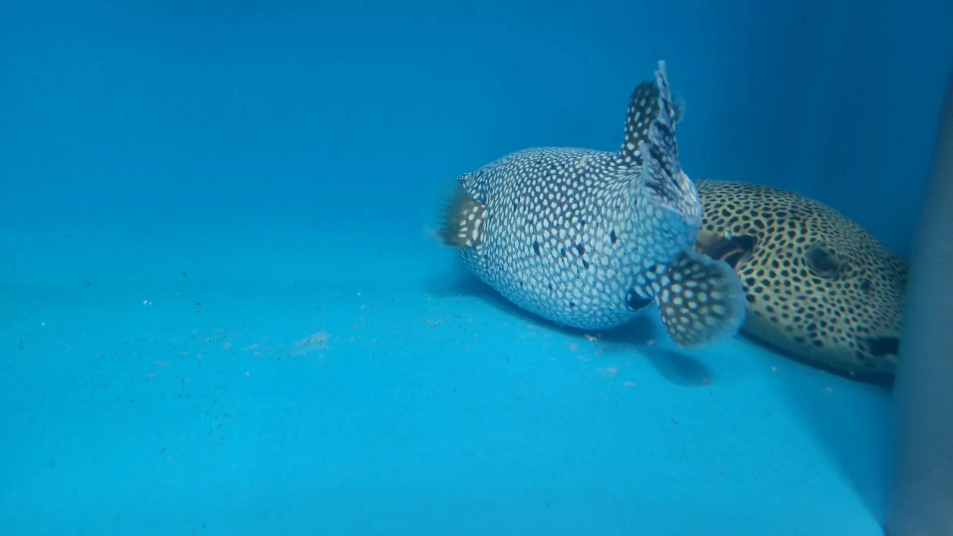
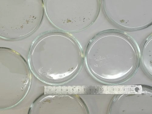
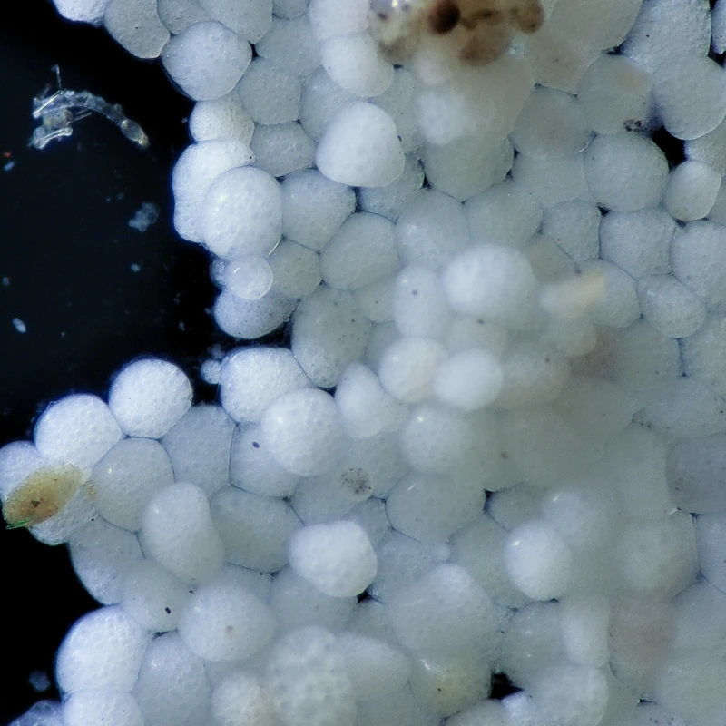
What has left the fish remains in the system. Petri dishes were placed on the bottom of an aquarium containing infected fish. Material accumulated on their surfaces that could easily have been mistaken for ordinary sediment or contamination. Under magnification, this material contained numerous attached stages of Cryptocaryon irritans.
Accumulations of attached stages
On the surfaces of the Petri dishes, the parasites occurred not only individually but also in dense multilayered accumulations. These areas formed films that, without magnification, could be mistaken for ordinary organic deposits.
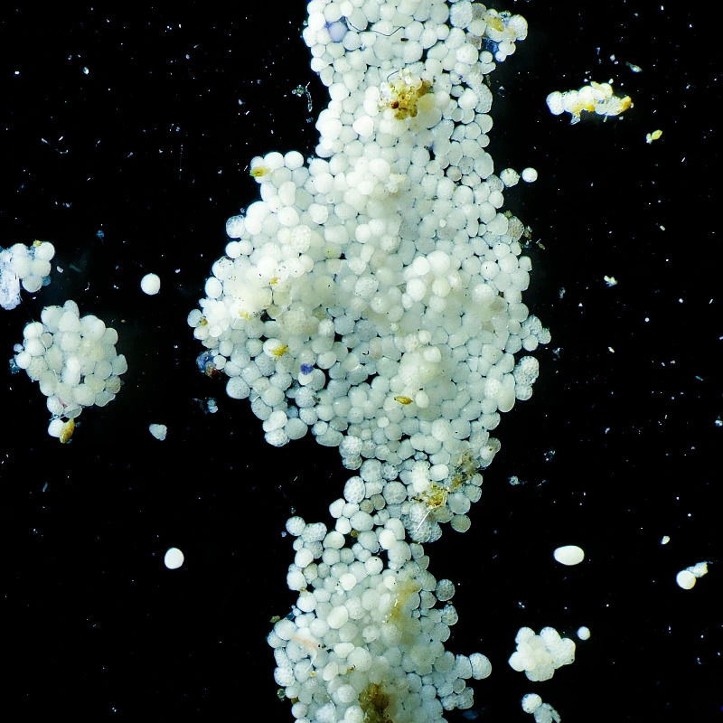
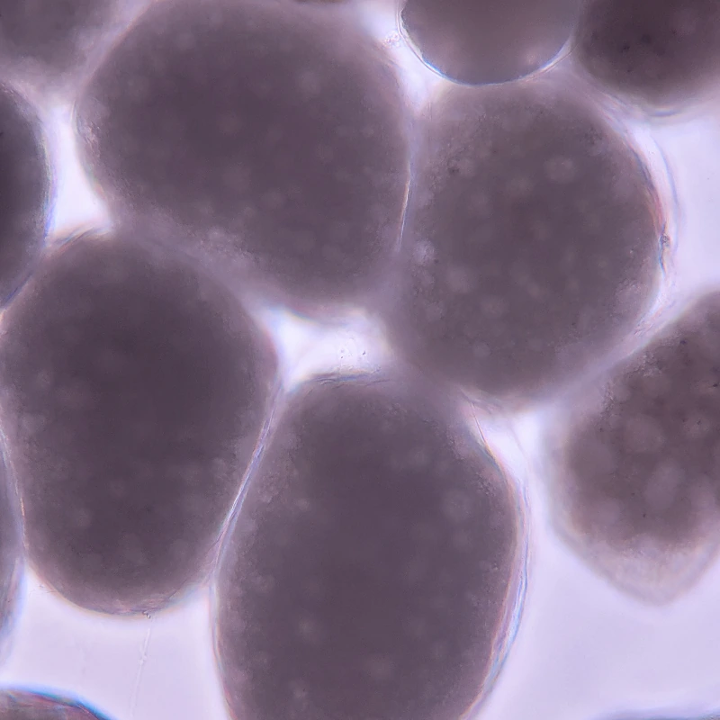
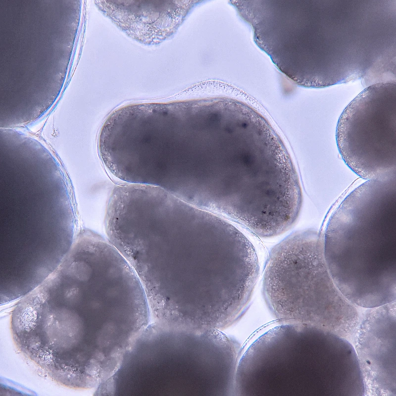
At higher magnification
When examined in transmitted light, rounded and oval structures surrounded by an outer wall are clearly visible. The internal contents of individual stages are heterogeneous, consistent with continuing development outside the host.
Parasites are removed together with mucus
When shedding of pathologically altered mucus was intensified, parasites left the fish surface together with dense mucus masses and cells of detached epithelium. They were present in large numbers in this material and often formed compact accumulations.
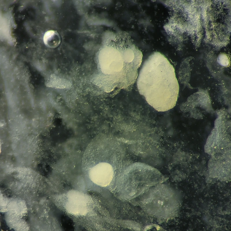
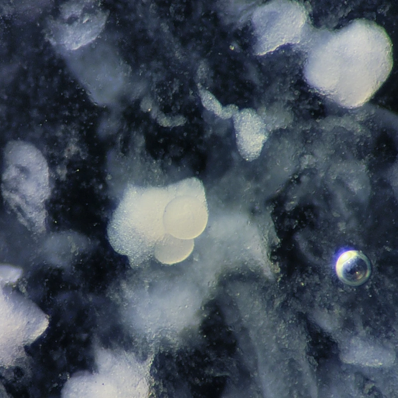
What appears externally to be mucus, sediment, or a film on a surface may contain large numbers of viable parasite stages.
Parasites that have left the fish do not disappear. Some remain in mucus and detached epithelium, while others attach to aquarium surfaces and continue developing outside the host. Temporary clearing of the skin therefore does not mean that the life cycle has stopped: unless the accumulated material is removed, the fish soon remains close to a new generation of infective stages (Burgess & Matthews, 1994).
These observations demonstrate a simple point: a decrease in the number of parasites on the fish does not mean that they have disappeared. A substantial part of the life cycle continues close to the host, within the same restricted system.
Action scheme: clean the fish and separate it from the parasite
If the fish is already heavily infected, treatment must address two objectives at the same time:
- rapidly reduce the number of parasites on the skin and gills;
- prevent the fish from immediately encountering the next generation of infective stages.
A treatment added to the water does not remove trophonts from the fish immediately. Its action is directed primarily at free-living stages of the parasite. In a severe case, waiting for treatment of the water to take effect may therefore be too slow.
First remove as much as possible of what is on the fish now. Then separate the fish from what remains in the system.
1. Direct treatment of the fish
When the species tolerates brief exposure to freshwater, this can be used to rinse the body surface and reduce the parasite burden.
The duration of the procedure must be determined by the condition and species-specific sensitivity of the fish. It should not be treated as a bath that the animal must endure at any cost. In some cases, a few minutes are sufficient to rinse the fish carefully while removing detached mucus and superficial pathological material.
In severe infestations, one procedure may not be sufficient. Several hours after the first treatment, intensified shedding of mucus and detached epithelium may begin. The fish can then be rinsed again to remove material that has already separated from the body surface.
More information: direct handling of fish and mechanical removal of external parasites
Simple Rules for Working with Aquarium Fish
Basic guidelines for diagnosing and caring for aquarium fish: what to do in case of mortality, how to interpret appetite, and when to treat …
The inability to remove every parasite in a single procedure is not a reason to leave all parasites on the fish.
2. Do not return the cleaned fish to accumulated parasite stages
After rinsing, the fish should be placed in a clean container. Returning it immediately to the same untreated aquarium restores contact with cysts, mucus, detached epithelium, and the infective stages emerging from them.
Two approaches are possible:
- transfer the fish to another prepared container;
- temporarily remove the fish, drain the aquarium completely, clean the aquarium and equipment, and then refill it.
The second option is particularly useful when only a small number of treatment tanks is available. The aquarium is then used not as a permanently contaminated environment, but as a container that can be repeatedly cleared of accumulated parasite stages.
3. Repeat transfer and cleaning
The more often the fish is separated from contaminated surfaces and newly emerged infective stages, the more strongly transmission of the parasite is disrupted (Colorni, 1985).
In theory, more frequent changes of container would be more effective than infrequent changes. In practice, every restraint, bath, and transfer imposes an additional burden on the fish. The frequency of procedures must therefore be chosen as a compromise between parasite removal and the animal’s tolerance of intervention.
For many fish, one complete treatment and transfer per day may already reduce reinfection substantially. The decision should take into account:
- severity of the condition;
- rate at which parasites reappear;
- tolerance of restraint and freshwater exposure;
- ability to maintain stable parameters in replacement containers;
- quality of aquarium cleaning between transfers.
Transfer works not because it “treats” the fish, but because it physically leaves part of the parasite’s life cycle behind.
4. Separate the fish from the bottom
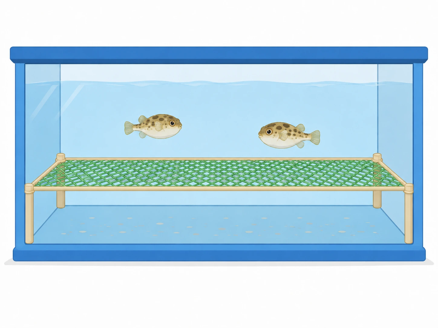
Theronts have limited motility: the maximum recorded upward swimming speed was 1.6 mm/s, and most theronts in the experiment were found within the lower five-centimetre layer of water. Increasing the distance between the fish and the bottom may therefore reduce the probability of reinfection (How et al., 2015).
A mesh platform can be installed above the bottom for this purpose. The mesh opening should be as large as possible while still preventing the fish from passing through. The mesh itself should have as little surface area as possible: thin structural elements reduce the area available for attachment of parasite stages.
The platform does not kill the parasite. It reduces the likelihood of direct contact between the fish and the surfaces on which the parasite accumulates and continues developing.
The purpose of the mesh is not to filter the water, but to prevent the fish from resting directly on a contaminated surface.
5. After stabilisation — treatment of the water
Physical removal of parasites and transfer to a clean container do not eliminate the need for further control of the system. Once the fish has stabilised, copper or another selected method can be used to target the free-living stages of the parasite.
The sequence, however, matters:
- first reduce the burden on the fish;
- remove accumulated material from the environment;
- limit reinfection;
- then target the remaining stages in the water.
Why “provoking emergence” with urea is questionable
Urea is sometimes proposed to stimulate the mass emergence of infective stages of Cryptocaryon irritans into the water, after which the released theronts are expected to be killed by a treatment. This scheme assumes that a large proportion of the parasites will enter the water simultaneously and be destroyed before they reach a host.
Urea has been investigated as an attractant for theronts that have already emerged and could potentially be used together with a trap, not as a proven means of synchronously inducing theront emergence from tomonts (Skilton et al., 2020).
A treatment concentration that is safe for the fish does not necessarily act instantaneously. If adding urea does increase the number of infective stages in the water, some may reach the fish and intensify infection before they are killed.
Such provocation is therefore meaningful only when it has been demonstrated that:
- the intervention really induces sufficiently synchronous emergence;
- the treatment kills the released stages rapidly enough;
- the effective concentration is maintained accurately;
- the fish is physically protected from contact with the released theronts.
When the fish can instead be transferred and the aquarium drained and cleaned, deliberately increasing the number of parasites in the water becomes an additional risk that is difficult to justify.
It is simpler to remove the fish from the parasite’s emergence zone than first to increase the number of infective stages and then attempt to kill them next to the host.
The main objective is to interrupt reinfection
The meaning of the entire scheme lies not in any single procedure, but in the sequential separation of fish and parasite. First, as many parasites as possible are removed from the body surface. The fish is then moved into clean water, the container is cleared of settled stages, and the animal is physically separated from the bottom where development of the parasite continues.
The objective is to make reinfection as unlikely as possible and the conditions for parasite persistence as unfavourable as possible.
The fewer parasites remain on the fish, the fewer viable stages persist in the environment, and the less often they reach the host again, the more time the animal gains for recovery. Subsequent treatment of the water then acts not against a continuously increasing infestation, but within a system in which the parasite’s life cycle has already been disrupted as much as possible.
References
-
Burgess P. J., Matthews R. A. 1994. Cryptocaryon irritans (Ciliophora): photoperiod and transmission in marine fish. Journal of the Marine Biological Association of the United Kingdom, 74(3): 535–542. https://doi.org/10.1017/S0025315400047652
-
Cervera L., González-Fernández C., Arizcun M., Cuesta A., Chaves-Pozo E. 2022. Severe natural outbreak of Cryptocaryon irritans in gilthead seabream produces leukocyte mobilization and innate immunity at the gill tissue. International Journal of Molecular Sciences, 23(2): 937. https://doi.org/10.3390/ijms23020937
-
Chi H., Goldstein M., Pichardo A., Wei Z.-H., Chang W.-J., Gong H. 2020. Infectivity and genes differentially expressed between young and aging theront cells of the marine fish parasite Cryptocaryon irritans. PLOS ONE, 15(8): e0238167. https://doi.org/10.1371/journal.pone.0238167
-
Colorni A. 1985. Aspects of the biology of Cryptocaryon irritans, and hyposalinity as a control measure in cultured gilthead sea bream Sparus aurata. Diseases of Aquatic Organisms, 1: 19–22. https://doi.org/10.3354/dao001019
-
Colorni A., Diamant A. 1993. Ultrastructural features of Cryptocaryon irritans, a ciliate parasite of marine fish. European Journal of Protistology, 29(4): 425–434. https://doi.org/10.1016/S0932-4739(11)80405-0
-
How K. H., Zenke K., Yoshinaga T. 2015. Dynamics and distribution properties of theronts of the parasitic ciliate Cryptocaryon irritans. Aquaculture, 438: 170–175. https://doi.org/10.1016/j.aquaculture.2014.12.013
-
Kaige N., Miyazaki T. 1985. A histopathological study of white spot disease in Japanese flounder. Fish Pathology, 20(1): 61–64. https://doi.org/10.3147/jsfp.20.61
-
Li Y., Jiang B., Mo Z., Li A., Dan X. 2022. Cryptocaryon irritans (Brown, 1951) is a serious threat to aquaculture of marine fish. Reviews in Aquaculture, 14(1): 218–236. https://doi.org/10.1111/raq.12594
-
Ma R., Ni B., Fan X., Warren A., Yin F., Gu F. 2016. Ultrastructure observation on the cells at different life history stages of Cryptocaryon irritans (Ciliophora: Prostomatea), a parasitic ciliate of marine fishes. Parasitology, 143(11): 1479–1489. https://doi.org/10.1017/S0031182016001074
-
Skilton D. C., Saunders R. J., Hutson K. S. 2020. Parasite attractants: identifying trap baits for parasite management in aquaculture. Aquaculture, 516: 734557. https://doi.org/10.1016/j.aquaculture.2019.734557
-
Yoshinaga T., Dickerson H. W. 1994. Laboratory propagation of Cryptocaryon irritans on a saltwater-adapted Poecilia hybrid, the black molly. Journal of Aquatic Animal Health, 6(3): 197–201. https://doi.org/10.1577/1548-8667%281994%29006%3C0197%3ALPOCIO%3E2.3.CO%3B2
-
Zec S., Hadfield C. A., Hungerford L. 2021. Retrospective review of copper sulfate immersion treatment in marine teleosts during quarantine at the National Aquarium of Baltimore from 2004 to 2016. Journal of Zoo and Wildlife Medicine, 52(1): 97–102. https://doi.org/10.1638/2020-0114
See also
Health Does Not Mean Sterility
Health Does Not Mean Sterility
Why the presence of microorganisms and parasites does not always mean disease, why immunity does not have to eliminate them completely, and …
Fossa Method: A Set of Principles
 Fossa Method
Observation, constraints, models, understanding, and the search for mechanisms. A collection of principles that gradually emerged from …
Fossa Method
Observation, constraints, models, understanding, and the search for mechanisms. A collection of principles that gradually emerged from …
Two Sides of the Same Dosage
 Two Sides of One Dosage
Aquarium medications: remedy or threat? Pros and cons of antibiotics, formalin, methylene blue, malachite green, and the FMC mixture.
Two Sides of One Dosage
Aquarium medications: remedy or threat? Pros and cons of antibiotics, formalin, methylene blue, malachite green, and the FMC mixture.
What to Do with a Fish After Arrival
 Initial Diagnostics After Fish Transportation
Microscopy, smears, staining methods, and common mistakes in early diagnostics.
Initial Diagnostics After Fish Transportation
Microscopy, smears, staining methods, and common mistakes in early diagnostics.