Method.
What remains of the Ziehl–Neelsen method if the method itself is changed?
Ziehl–Neelsen staining is used to detect acid-fast bacteria, especially mycobacteria (World Health Organization, 1982). In the classical procedure, the preparation is stained with carbol fuchsin with heating, then decolorized with acid or acid-alcohol and counterstained with a contrasting dye (World Health Organization, 1982) (World Health Organization, 1999).
The logic of the method is simple. The mycobacterial cell envelope is rich in lipids and mycolic acids and stains poorly with ordinary aqueous dyes (Caulfield & Wengenack, 2016). Carbol fuchsin in the presence of phenol and heat makes it possible to achieve persistent staining of these structures (Caulfield & Wengenack, 2016) (World Health Organization, 1982). After acid treatment, ordinary tissues and non-acid-fast bacteria lose the fuchsin, while acid-fast structures remain red (Caulfield & Wengenack, 2016). Counterstaining then makes the background blue or green. As a result, we obtain a very convenient picture: red rods against a contrasting background. This is usually where the description of the method ends.
But this is precisely where a more interesting question begins: “Which parts of this sequence are actually necessary for our task?”
The method and the task are not the same thing
If the task is “to demonstrate acid-fastness of bacteria”, everything is unambiguous: the acid decolorization step is necessary by definition. Without it, we simply do not test the property by which the bacteria are called acid-fast. But in routine work, the task may be different.
For example: to see characteristic mycobacteria-like rods in tissue material, find suspicious clusters, and decide whether the material should be investigated further.
That is no longer quite the same question. The Ziehl–Neelsen method is a specific sequence of reagents and procedures (as is any method). If phenol or acid is removed, or the heating regime is changed, strictly speaking this is no longer classical Ziehl–Neelsen staining. But that does not mean that the resulting variant automatically becomes useless. It simply has to be called something else and evaluated according to a different task.
What if phenol is removed?
Phenol in the classical method is not needed “because the recipe says so”. It is part of classical carbol fuchsin, while heating is used to obtain intense staining of mycobacteria (World Health Organization, 1999) (World Health Organization, 1982). At the same time, phenol-free modifications have been developed and their diagnostic suitability has been evaluated (Ramos et al., 2019). And here a practical question arises: how essential is this particular classical combination of factors? Phenol is a toxic and unpleasant reagent to work with. If it is unavailable or undesirable to use, there is no need to pretend that we are still performing Ziehl–Neelsen staining.
We can ask a different question:
Can sufficient penetration of fuchsin be achieved in another way?
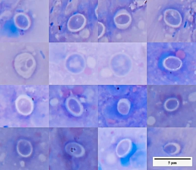
In my preparations, intensive heating of fuchsin without phenol produced clearly visible red rod-shaped structures. This does not prove that phenol and heating are fully interchangeable, and it does not automatically turn such staining into a new Ziehl–Neelsen variant. But it shows that, for a specific practical task, the function of one component of the classical procedure can be partly compensated by another treatment.
This is where an important boundary lies. The method can be changed. What cannot be done is to pretend afterward that the method was not changed.
If the classical procedure has been modified, that should be stated explicitly, together with a description of what exactly was done.
Is acid always necessary?
With acid, the situation is even clearer. In classical staining, acid decolorization creates the main diagnostic criterion: some structures lose fuchsin, while acid-fast ones retain it. Therefore, without this step one cannot claim: “Acid-fast bacteria were detected”.
Acid-fastness was simply not tested in that case. At the same time, the acid itself is not some uniquely prescribed reagent. Different versions of the method use different acids and concentrations for differentiation: for example, 15–25% sulfuric acid or acid-alcohol containing HCl (World Health Organization, 1982) (World Health Organization, 1999). WHO materials also note that hydrochloric, sulfuric, and nitric acids have been used as decolorizers (World Health Organization, n.d.). In other words, the diagnostic meaning lies not in the name of a particular acid, but in the fact that sufficiently strong decolorization is performed, after which the bacteria retain fuchsin.
Try decolorizing with a concentrated citric acid solution — and see whether the name of the acid is really the decisive factor if previously stained structures lose their color and become almost transparent.
Important: this suggestion is an experimental test of the function of the step, not a reference to a validated Ziehl–Neelsen staining protocol. In the standard guidelines reviewed, citric acid is not listed as a routine decolorizer.
Therefore, concentration should also be viewed not as a sacred number from a recipe, but as part of the differentiation conditions. Treatment that is too weak will leave fuchsin where it should have been removed…treatment that is too harsh may impair staining even in genuinely acid-fast bacteria. But if the task is not to demonstrate acid-fastness, but to visualize suspicious microorganisms, acid ceases to be an essential part of that particular task.
After intensive staining with carbol fuchsin, the preparation can be counterstained with a contrasting dye. In my case, a Löffler counterstain was used. Ordinary dyes without phenol and heating penetrate the lipid-rich cell envelope much less effectively, so rods that have already taken up fuchsin remain bright red, while surrounding tissues and the background stain blue.
This contrast makes them easy to distinguish, but by itself still does not demonstrate acid-fastness. Such a preparation is not the result of a complete differential acid-fast stain. It shows the presence and morphology of stained bacteria and allows one to decide whether there are grounds to perform a full test of acid-fastness.
Why morphology matters here
Mycobacteria-like bacteria in such preparations have a fairly characteristic appearance: thin red rods, isolated or arranged in small groups and clusters, sometimes with uneven staining. The appearance of a bacterium alone, of course, does not determine its genus, let alone its species. But in routine diagnostics, we almost never rely on a single feature. We assess several things at once:
- bacterial shape and size;
- their arrangement;
- staining;
- tissue context;
- the presence of granulomatous changes;
- reproducibility of the finding in different areas of the material.
Therefore, characteristic red rods in an expected morphological context can be a sufficiently strong reason to continue the investigation using classical acid-fast staining, culture, PCR, or another method.
Where are the “traditions instead of science” here?
The problem begins when a sequence of actions stops being regarded as a way to solve a task and turns into a ritual. Phenol is needed because “Ziehl–Neelsen uses phenol”. Acid is needed because “that is how the method is supposed to be done”. Heating must last exactly that long because “that is what is written”. But every step has a function.
And if we understand that function, it becomes possible to ask sensible questions:
- What exactly are we trying to detect?
- Which step of the method creates the diagnostic feature we need?
- Can that step be changed?
- What will we lose after changing it?
- Which conclusions remain valid afterward, and which no longer do?
Rejecting a traditional protocol does not mean rejecting science. Quite the opposite: a scientific approach begins when we stop treating a method as an immutable set of actions and start understanding why each action is performed.
But there is another side to this:
If we have moved beyond the boundaries of the standard method, we can no longer hide behind its name.
It is acceptable to say: “A modified carbol fuchsin stain without acid decolorization was used. Intensely stained rod-shaped microorganisms were detected.”
It is not acceptable to say: “Ziehl–Neelsen staining was performed; acid-fast bacteria were detected.”
These are already different statements.
Conclusion
A method can be changed. But when the method changes, the boundaries of permissible conclusions change with it.
Sources
World Health Organization, 1982. Staining methods for acid-fast bacteria.
Classical Ziehl–Neelsen procedure with heating of carbol fuchsin and decolorization with 15% sulfuric acid.
https://iris.who.int/bitstream/handle/10665/260733/PMC2366253.pdf?isAllowed=y&sequence=1
World Health Organization, 1999. Tuberculosis: a manual for medical students. WHO/CDS/TB/99.272.
Describes hot Ziehl–Neelsen staining, acid-alcohol decolorization, and a variant using 25% sulfuric acid.
https://iris.who.int/bitstream/handle/10665/68559/WHO_CDS_TB_99.272.pdf
Caulfield, A.J., Wengenack, N.L., 2016. Diagnosis of active tuberculosis disease: From microscopy to molecular techniques. Journal of Clinical Tuberculosis and Other Mycobacterial Diseases, 4, 33–43.
Review of acid-fast staining, the hydrophobic mycobacterial cell wall, mycolic acids, and resistance of the dye complex to decolorization.
https://pmc.ncbi.nlm.nih.gov/articles/PMC6850262/
World Health Organization, 1982. General tests / staining procedure for acid-fast bacilli.
Notes that the high lipid content impedes dye penetration; prolonged exposure to carbol fuchsin or heating is used to achieve staining.
https://iris.who.int/bitstream/10665/62463/1/BLG_UNDP_82.1_eng.pdf
Ramos, A. et al., 2019. GASMoC method: a phenol-free technique to detect acid-fast bacilli.
An example of a study evaluating a modified phenol-free technique for detecting acid-fast bacteria.
https://pubmed.ncbi.nlm.nih.gov/31654127/
World Health Organization, n.d. Ziehl–Neelsen procedure: fundamentals.
The document explicitly states that different acids, including hydrochloric, sulfuric, and nitric acid, may be used for decolorization; it also describes the acid step as a form of regressive differentiation.
https://applications.emro.who.int/docs/EM_SND_MTG_STR_LEP_CNT_9_3_EN.pdf
Cambau, E. et al., 2024. Acid-Fast Positive and Acid-Fast Negative Mycobacterium tuberculosis: The Koch Paradox.
Historical review of the development of the method: phenol introduced by Ziehl, heating by Rindfleisch, and 25% sulfuric acid by Neelsen; a useful demonstration that the “classical” procedure itself emerged through successive modifications.
https://pmc.ncbi.nlm.nih.gov/articles/PMC11687472/
See also:
Scientific Method. A Practical Guide.
 Scientific Method. A Practical Guide.
How knowledge is formed, why confidence often outpaces understanding, and how the scientific method helps distinguish observation from …
Scientific Method. A Practical Guide.
How knowledge is formed, why confidence often outpaces understanding, and how the scientific method helps distinguish observation from …
Ichthyophthiriosis Without Myths
 Ichthyophthiriosis Without Myths
A science-based analysis of Ichthyophthirius multifiliis: life cycle, cyst terminology, the myth of parasite ubiquity, and real mechanisms …
Ichthyophthiriosis Without Myths
A science-based analysis of Ichthyophthirius multifiliis: life cycle, cyst terminology, the myth of parasite ubiquity, and real mechanisms …
Hexamitids Without Myths
 Hexamitids Without Myths
Why hexamitids are not a normal component of fish intestinal microflora, where the myth of 'internal parasites' comes from, and how …
Hexamitids Without Myths
Why hexamitids are not a normal component of fish intestinal microflora, where the myth of 'internal parasites' comes from, and how …
Initial Diagnostics After Fish Transportation
 Initial Diagnostics After Fish Transportation
Microscopy, smears, staining methods, and common mistakes in early diagnostics.
Initial Diagnostics After Fish Transportation
Microscopy, smears, staining methods, and common mistakes in early diagnostics.