- In most cases, the disease is caused by supersaturation of water with nitrogen, less commonly oxygen. This typically happens when water is heated in closed systems where gases cannot freely escape — such as heat exchangers in power plants or boilers in hatcheries.
- Air bubbles in the water, on glass, or on plants are dangerous — they can block the gills of fish and cause death.
- External signs of gas embolism appear a few hours after fish are placed in supersaturated water. The higher the gas saturation level, the faster the symptoms develop and the sooner the fish may die. At lower saturation levels, fish may survive longer, and signs like the location and shape of bubbles become more variable.
- During necropsy of larger fish, foamy blood may emerge from blood vessels. Other signs include: hyperactivity, hypersensitivity to mechanical stimuli, loss of balance, twitching fins, scale erection, ray separation, corneal opacity, lightening or darkening of body coloration, loss of vision, exophthalmia.
- Causes may include: excessive oxygen production by plants, overly aggressive artificial aeration, or the use of untreated tap water. The disease develops in conditions of oxygen and nitrogen oversaturation.
- Oxygen entering the bloodstream in excess forms tiny bubbles that block blood vessels. When bubbles enter capillaries, they are often larger than the vessel’s diameter, and the heart is not strong enough to push them through — resulting in blockage.
The diagnosis is based on clinical signs, necropsy findings, and water analysis, specifically the level of dissolved oxygen.
Scientific Perspective
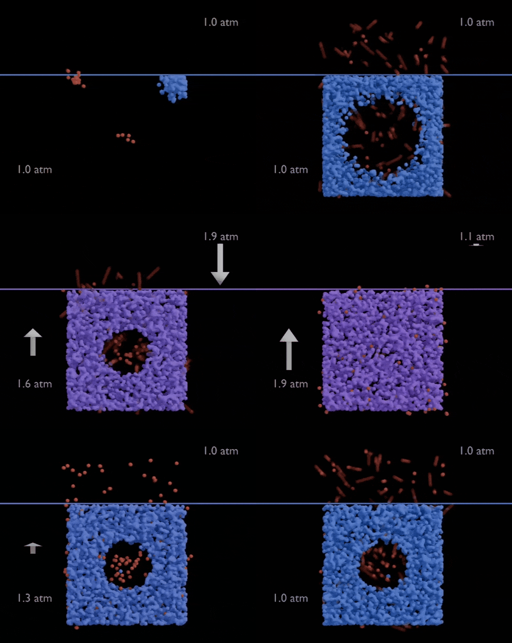
Under increased pressure, bubbles do not form. Gas molecules remain dissolved, mixing with water molecules.
Gas Bubble Disease (GBD) is a pathological condition in which free gas bubbles form within the blood and tissues of fish. These bubbles impair circulation, damage the gills and internal organs, cause tissue ischemia, and may ultimately lead to death.
At first glance, the explanation seems obvious: if bubbles are present in the water, they must enter the fish and cause the disease.
This is exactly how gas bubble disease is described in many articles, books, and even textbooks.
However, this explanation is incorrect.
Gas bubble disease does not develop because fish come into contact with air bubbles. It develops because the water contains an excessive amount of dissolved gases. These are fundamentally different phenomena.
The bubbles visible in an aquarium and the bubbles forming inside a fish’s blood vessels have completely different origins.
What causes Gas Bubble Disease?
The primary cause of GBD is gas supersaturation.
Under certain conditions, water can dissolve more gas than would normally exist at equilibrium for a given pressure and temperature. In most cases, the excess consists mainly of nitrogen, less commonly oxygen or a mixture of atmospheric gases.
As long as pressure remains elevated, the excess gas may remain completely dissolved. The water appears perfectly clear, even though it actually contains more dissolved gas than equilibrium allows.
This is the dangerous condition.
When water contains excess dissolved gases, the partial pressure of these gases at the gill surface becomes higher than in the blood. A diffusion gradient is established, allowing gases to diffuse across the gill epithelium until a new equilibrium is reached.
This is how gas bubble disease develops.
Where do the bubbles form?
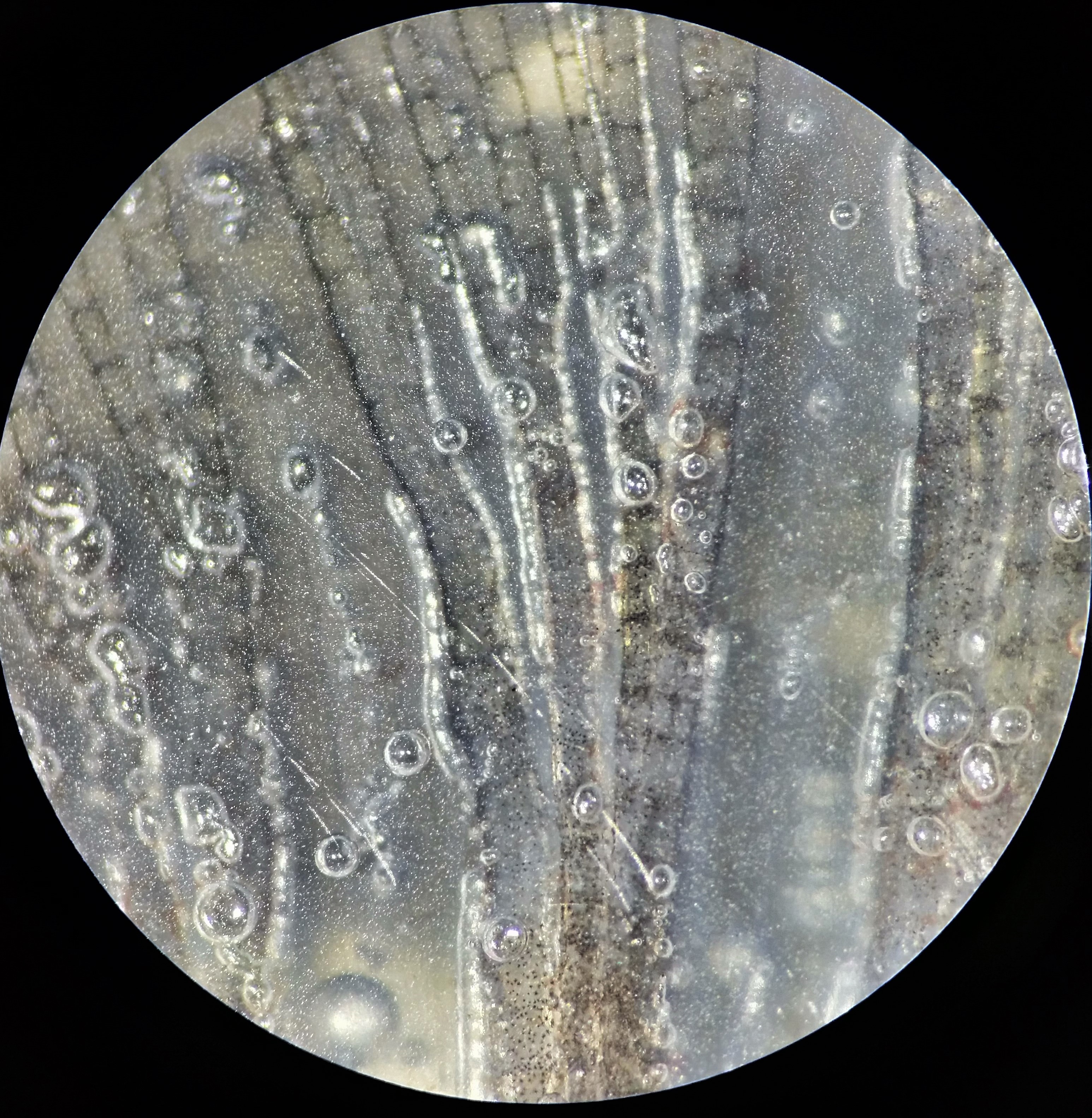
Elongated gas bubbles within the fin blood vessels. Their characteristic shape indicates formation inside the bloodstream under pressure.
This is one of the most important questions.
Gas bubbles do not enter the fish from the surrounding water.
Instead, they form inside the body, when dissolved gases can no longer remain in solution within the blood and tissue fluids.
Therefore, finding bubbles inside blood vessels does not mean they entered through the gills. They formed exactly where dissolved gas came out of solution.
The same physical mechanism is well known outside ichthyopathology. It is responsible for decompression sickness in divers.
Why may there be no visible bubbles at all?
This is where the most common misconception begins.
Most people judge the risk by the number of bubbles visible in the water.
In reality, the opposite is often true.
Under elevated pressure, water can hold a large amount of dissolved gas without forming bubbles. The gas remains completely dissolved and therefore invisible.
When pressure decreases, gas solubility decreases as well, and the excess gas begins to leave the solution as visible bubbles.
In other words, visible bubbles often indicate not the beginning of the problem, but that the water has already started releasing excess dissolved gas.
Pressure keeps gases dissolved
Pressure affects gas solubility.
This process is described by Henry’s Law.
The amount of gas that water can hold is determined by its partial pressure.
C = kH × P
where:
C — concentration of dissolved gas;
kH — Henry’s constant;
P — partial pressure of the gas.
The practical implication is straightforward.
Increasing pressure allows water to dissolve more gas.
When pressure subsequently decreases, gas solubility falls, and the excess gas begins to leave the solution.
This is why elevated pressure within water treatment systems can create conditions for gas supersaturation, whereas visible bubbles often appear only after pressure has decreased.
Why is this important?
Preventing gas bubble disease is impossible if assessment is based solely on visible bubbles.
Proper preventive measures include:
controlling pressure within the water treatment system;
monitoring Total Gas Pressure (TGP), whenever possible;
removing excess dissolved gas by aeration or cascade degassing.
During necropsy
The presence of gas within tissues during necropsy is not, by itself, evidence of gas bubble disease. Postmortem gas formation is a common consequence of carcass warming and autolysis. As temperature rises, gas solubility decreases, allowing dissolved gases to leave solution and accumulate within blood vessels and beneath serosal membranes.
Therefore, gas observed during necropsy may reflect not only processes that occurred during life, but also postmortem changes.
Practical observation
Based on our observations, conventional aquarium pumps rarely generate a level of gas supersaturation sufficient to produce classical gas bubble disease by themselves.
Even with air entrainment and pressure increases of approximately 0.5 bar above atmospheric pressure, the increase in gas solubility remains limited and is generally not associated with the characteristic clinical signs of the disease.
Risk arises when several factors occur simultaneously:
air entering the pressurized pipeline;
gas accumulation under elevated pressure;
rapid pressure reduction (decompression).
Only the combination of these factors is likely to produce hazardous gas supersaturation.
Conclusion
Gas bubble disease does not develop because bubbles enter the fish.
It develops because the water contains an excess of dissolved gases.
The bubbles visible in an aquarium are more often a consequence of degassing than the cause of the disease.
Therefore, diagnosis should focus not on the bubbles themselves, but on identifying the conditions that lead to gas supersaturation.
REFERENCES
Legendre D, Zenit R. Gas bubble dynamics. Published online January 6, 2025. doi:10.48550/arXiv.2501.02988
Chen X, Zhang Z, Qin Y, et al. An in-situ study in the Xijiang River basin revealed adverse effects of total dissolved gas supersaturation on fish. Ecotoxicol Environ Saf. 2025;289:117663. doi:10.1016/j.ecoenv.2024.117663
Hong WH, Choi JY, Cho HS, et al. Gas bubble disease in captive Golden Trevally: Pathological insights and needs for life support system and water quality management. J Aquat Anim Health. 2024;36(4):348-354. doi:10.1002/aah.10237
Máchová J, Faina R, Randak T, et al. Fish death caused by gas bubble disease: A case report. Veterinární Medicína. 2017;62:231-237. doi:10.17221/153/2016-VETMED
Pleizier NK, Brauner CJ. Causes and consequences of gas bubble trauma on fish gill function. J Comp Physiol B. 2024;194(5):739-747. doi:10.1007/s00360-024-01538-4
Wang H, Wang Y, Li K, Liang R, Zhao W. Tolerance threshold of a pelagic species in China to total dissolved gas supersaturation: from the perspective of survival characteristics and swimming ability. Conserv Physiol. 2024;12(1):coae023. doi:10.1093/conphys/coae023
Part II. Consequences.
Gas Bubble Disease. Part II. Consequences.Comparison of normal and pathological findings in fish necropsy: liver, kidney, gills, and brain. How to distinguish acceptable variation …
---
FAQ
Are visible bubbles dangerous?
Not necessarily. They often indicate gas release, not accumulation.
Why do fish die then?
Because of dissolved gas supersaturation, not visible bubbles.
Can bubbles enter the bloodstream from water?
No. They form inside tissues.
Can the problem be detected visually?
Not always. Supersaturation may be invisible.
Does cold water cause the disease?
No. The key factor is gas supersaturation.
See also:
Scientific Method. A Practical Guide.
Scientific Method. A Practical Guide.How knowledge is formed, why confidence often outpaces understanding, and how the scientific method helps distinguish observation from …
Granulomas and intracellular agents in fish.
Granulomas and intracellular agents in fishA collection of real observations: intracellular yeast-like organisms, coccidia, cryptosporidia, and fungal structures in fish.
Cryptocaryon irritans: Not Just White Spots.
Cryptocaryon irritans: Not Just White SpotsCryptocaryon irritans. is a ciliated protozoan parasite, often visible to the naked eye. Tangs, butterflyfish, and boxfish are particularly …
 Gas Bubble Disease. Part II. Consequences.
Comparison of normal and pathological findings in fish necropsy: liver, kidney, gills, and brain. How to distinguish acceptable variation …
Gas Bubble Disease. Part II. Consequences.
Comparison of normal and pathological findings in fish necropsy: liver, kidney, gills, and brain. How to distinguish acceptable variation …
 Scientific Method. A Practical Guide.
How knowledge is formed, why confidence often outpaces understanding, and how the scientific method helps distinguish observation from …
Scientific Method. A Practical Guide.
How knowledge is formed, why confidence often outpaces understanding, and how the scientific method helps distinguish observation from …
 Granulomas and intracellular agents in fish
A collection of real observations: intracellular yeast-like organisms, coccidia, cryptosporidia, and fungal structures in fish.
Granulomas and intracellular agents in fish
A collection of real observations: intracellular yeast-like organisms, coccidia, cryptosporidia, and fungal structures in fish.
 Cryptocaryon irritans: Not Just White Spots
Cryptocaryon irritans. is a ciliated protozoan parasite, often visible to the naked eye. Tangs, butterflyfish, and boxfish are particularly …
Cryptocaryon irritans: Not Just White Spots
Cryptocaryon irritans. is a ciliated protozoan parasite, often visible to the naked eye. Tangs, butterflyfish, and boxfish are particularly …