Granulomatous lesions in fish should not be automatically attributed to mycobacteriosis.
A similar gross presentation may reflect completely different etiologies, and microscopy is often decisive.
This page presents selected observations and morphological examples.
Each card links to a separate page with a full analysis.
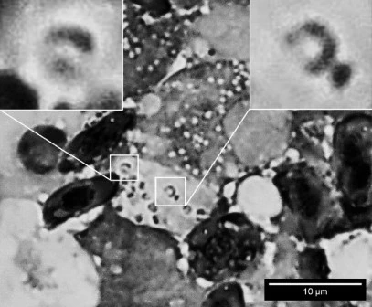
Histoplasma-like structures
Small intracellular yeast-like cells (1–3 µm) within histiocytes of the spleen and kidney. Halo, budding, and horseshoe-like basophilic structures.

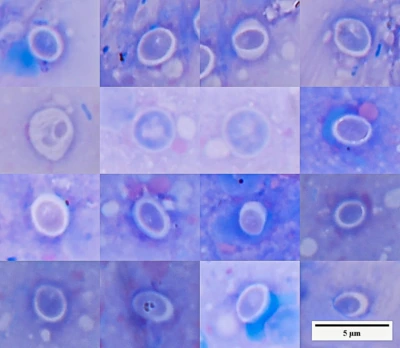
Cryptosporidium-like structures
Small uniform round structures with a distinct halo. Morphologically similar to cryptosporidia but require cautious interpretation.
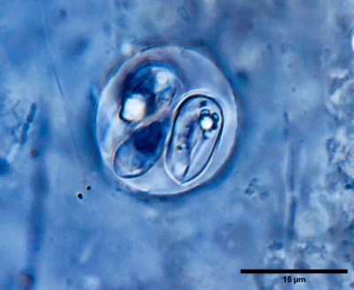
Goussia
Coccidia with oval oocysts and elongated sporozoites inside sporocysts. A characteristic example of intestinal parasites in fish.
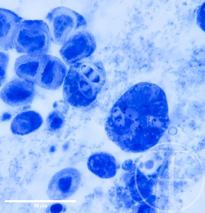
Intracellular fungal forms
Large cells with multiple inclusions and tissue-associated forms potentially linked to fungal etiology.
Important
In pathology and microbiology, a granuloma is a non-specific response.
It indicates a chronic inflammatory process but does not identify the causative agent by itself.
FAQ
Are granulomas always caused by mycobacteria?
No. Granulomas are a non-specific response with multiple possible causes.
Can diagnosis be made by gross appearance?
No. Similar lesions may have completely different origins.
Why is microscopy important?
It reveals intracellular structures and helps differentiate processes.
Are all parasite-like structures actually parasites?
No. Morphology alone can be misleading.
Why avoid quick conclusions?
Because identical patterns can arise from different mechanisms.