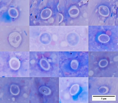
“Granulomas? Then it must be fish tuberculosis”
When granulomas are found in the parenchymal organs of a fish, the next thought often comes almost automatically:
Mycobacteriosis.
Or, more simply:
Fish tuberculosis.
The association is understandable. Granulomas are indeed very common in fish with mycobacteriosis. The problem begins when a characteristic response of the organism is treated as the name of the causative agent. First, we need to establish what we actually saw.
What is a granuloma?
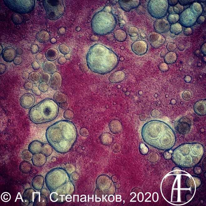
One classical definition of a granuloma is a compact, organized collection of mature mononuclear phagocytes, primarily macrophages (large immune cells that engulf bacteria, viruses, and cellular debris).
Necrosis (irreversible cell death), giant cells (very large, usually multinucleated cells), lymphocytes (a type of immune cell), and other familiar components of granulomas may be present, but none is essential to the definition. What matters is the organized accumulation of mature macrophages
(Pagán & Ramakrishnan, 2018).
A simpler way to express the modern definition is:
A granuloma is an organized attempt by macrophages to deal with something that a single macrophage could not handle.
Pagán and Ramakrishnan offer a particularly clear model:
-
If the first phagocyte engulfs an object and manages to destroy it, there is no need for a granuloma.
-
If the object persists, is difficult to break down, or is too large, additional cells are recruited and a more organized structure forms (Pagán & Ramakrishnan, 2018).
In other words, the very appearance of a granuloma tells us primarily that:
something was persistent enough that an ordinary phagocytic response (an immune defense in which cells called phagocytes, such as macrophages, recognize, engulf, and digest foreign particles, bacteria, and dead cells) was insufficient.
It does not even have to be “foreign”
As early as 1925, Margaret Reed Lewis observed the behavior of monocytes in culture. These cells responded not only to tuberculosis bacteria or experimentally introduced coal particles.
They also engulfed:
- the body’s own melanin granules (small intracellular compartments, or organelles, that contain the natural dark pigment melanin);
- dead granulocytes (a type of white blood cell containing prominent granules with biologically active substances involved in immune defense and inflammation);
- erythrocytes (red blood cells that transport oxygen from the respiratory organs to the tissues and carry carbon dioxide back).
During cultivation, monocytes developed into macrophages (large immune cells that engulf bacteria, viruses, and cellular debris). After several days, cells that had engulfed difficult-to-remove material acquired an epithelioid appearance, and some fused to form multinucleated giant cells (Pagán & Ramakrishnan, 2018).
Thus, the distinction between “self” and “foreign” is not the only thing that matters in the development of a granulomatous response.
Another question is more important:
Can the material be removed quickly, or not?
A dead cell does not literally become a “foreign body.” It remains material belonging to the organism. But when a cell breaks down, it leaves behind membranes (thin, flexible layers of lipids and proteins that enclosed the cell and its internal organelles, separating them from the surroundings), lipids (a broad group of fats and fat-like organic substances that are insoluble in water but soluble in organic solvents), proteins (complex organic molecules made of amino-acid chains that serve as structural components and carry out many biological functions), and other cellular debris (the remains of destroyed cells and tissues after cell death, inflammation, or injury). This material has to be cleared away. If it persists in the tissues, phagocytes (including macrophages, monocytes, and neutrophils) continue to respond, attempting to engulf and remove the remnants of destroyed cells. Such a response can become prolonged and contribute to chronic granulomatous inflammation.
That is why the equation:
“granuloma = infectious agent”
is too simplistic from the outset.
A mechanism older than fish
The granulomatous response appears to be much older than one might expect of the complex cellular immune system of vertebrates. Granulomas are formed not only by mammals, birds, and fish. They have also been described in mollusks, worms, and insects (Pagán & Ramakrishnan, 2018). One example is particularly striking. A parasitoid wasp lays an egg in a fruit-fly larva. The egg is too large for an individual phagocyte to engulf. Instead, cells surround it and form an organized granuloma-like structure. The underlying task remains the same:
If you cannot destroy it, isolate it.
In the freshwater clam Corbicula fluminea, a granulomatous response can even develop around the animal’s own larvae when, for some reason, they are not released normally. Their remnants are subsequently resorbed (Pagán & Ramakrishnan, 2018). It is hard to imagine a clearer example of why the presence of a granuloma alone tells us nothing specific about mycobacteria.
Many different causes
A granulomatous response may be triggered by very different agents. In their review, Pagán and Ramakrishnan list bacteria, fungi, protozoa, helminths, and viruses among the infectious causes. Granulomas may also arise around foreign material and in a variety of noninfectious inflammatory conditions (Pagán & Ramakrishnan, 2018).
So this line of reasoning:
granuloma → mycobacterium
does not work.
A better formulation is:
granuloma → a persistent factor → determine what that factor is.
In aquarium practice, mycobacteriosis is indeed among the first possibilities to investigate when multiple granulomas are present. But the leading differential diagnosis is not yet an established diagnosis.
Mycobacteria
The story becomes even more interesting with mycobacteria. For a long time, a granuloma was imagined almost as a fortress built by the organism around a bacterium:
Here are the bacteria; here is a wall of immune cells around them; the infection has been contained.
This mechanism does exist. But it turned out to be only part of the picture. Studies of Mycobacterium marinum in zebrafish have shown that pathogenic mycobacteria can exploit the very process of granuloma formation:
An infected macrophage attracts new macrophages. It dies. Another macrophage engulfs its remains, together with the bacteria inside. Now the next macrophage is infected. It, too, becomes part of the granuloma. In these experiments, the number of infected macrophages began to rise sharply after cellular aggregates formed. At the same time, the number of viable mycobacteria increased (Pagán & Ramakrishnan, 2015).
The result is almost paradoxical:
the organism recruits cells to contain the infection, while the bacterium uses the arriving cells as new sites for replication.
Nor is a mature granuloma necessarily a sealed capsule. Cells continue to enter and leave it, and infected macrophages can move away from the primary focus and contribute to the formation of new lesions (Pagán & Ramakrishnan, 2015).
That makes the modern view of granulomas far more interesting than the old one:
A granuloma can both contain an infection and help the pathogen spread.
Which process predominates depends on the stage of infection, the state of the immune response, and the properties of the pathogen itself.
Many granulomas — many bacteria?
Not necessarily.
A granuloma reflects more than the number of pathogens present.
It reflects how the organism interacts with them.
When the cellular response is effectively organized, well-defined granulomas may develop.
When macrophage function is impaired, lesions may become disorganized and necrotic even while the bacterial burden is very high.
In experimental models where tumor necrosis factor (TNF)—a signaling protein through which immune cells regulate inflammation and defense against infection—was disrupted, mycobacteria proliferated actively while granulomas lost their organized structure
(Pagán & Ramakrishnan, 2015).
Thus, “generalized mycobacteriosis, but very few well-formed granulomas” is not a contradiction at all. Sometimes it may indicate the opposite of what one would expect: the immune system is failing to contain the infection effectively.
What if we use Ziehl–Neelsen staining?
This is where we move from a suspicion to the next level of evidence.
If numerous rod-shaped bacteria are found within granulomas and retain carbol fuchsin after acid decolorization, the hypothesis of mycobacteriosis becomes considerably stronger. But here, too, we must avoid skipping another step. Ziehl–Neelsen staining demonstrates acid-fastness. It does not automatically establish the species of bacterium.
More on this: Traditions Instead of Science I. Method: Staining and the Limits of Interpretation
Species-level identification requires other methods, such as culture, PCR, sequencing, or a combination of these approaches.
A granuloma tells us:
An organized chronic tissue response is taking place.
Staining tells us:
Structures with particular staining properties are present here.
Molecular or culture-based methods bring us closer to answering:
Who exactly is present?
One does not replace the other.
Sometimes a granuloma has nothing to do with bacteria
Here are two fish spleens with numerous whitish lesions. In the first case, mycobacteria were detected by microscopy. In the second, intracellular microorganisms morphologically resembling Histoplasma were found.
The first photograph is included specifically for comparison. The two organs show similar changes to the naked eye, but microscopy reveals very different findings. This is precisely why numerous whitish nodules do not, by themselves, identify the cause.
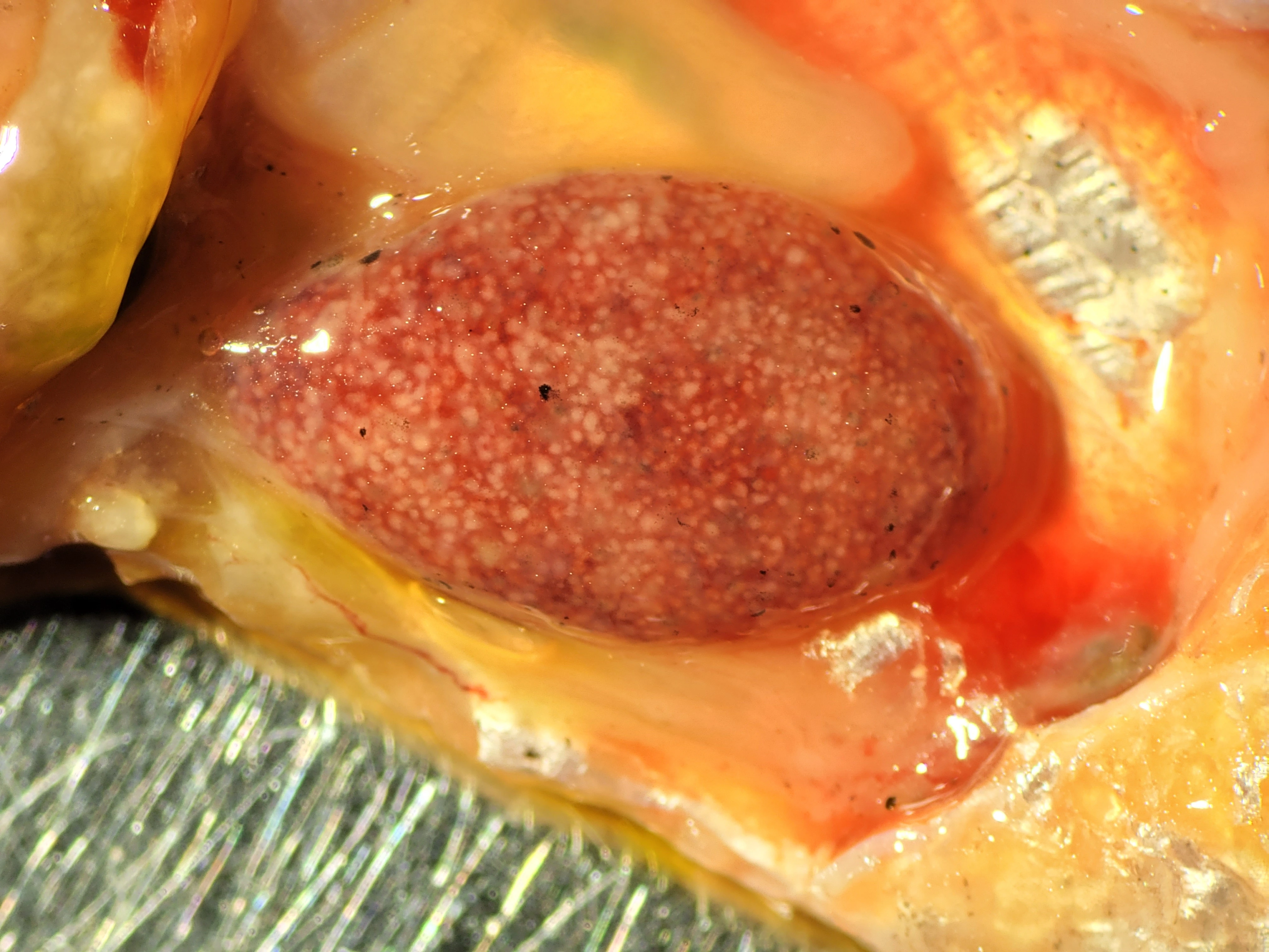
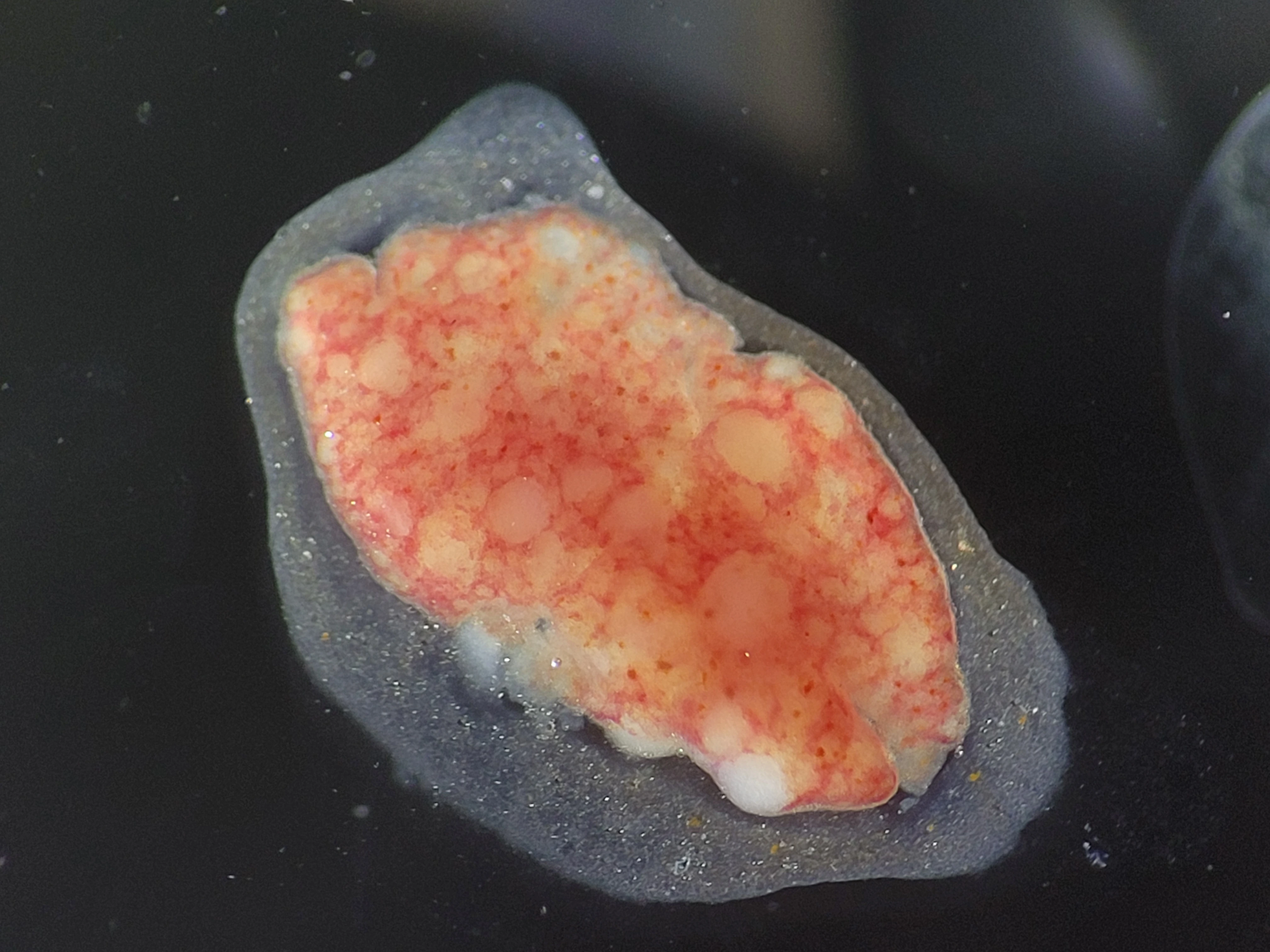
It is possible to find numerous granulomatous lesions without detecting acid-fast bacteria in them. The cause may be an entirely different process—for example, a parasitic infection. That is why it is better to describe what is actually visible first:
“multiple granulomas”
and only then investigate their etiology.
Because the entry:
“multiple granulomas — mycobacteriosis”
quietly turns an observation into a diagnosis.
Granuloma shape matters too
Liver of Apteronotus albifrons with granulomatous lesions (acid-fast bacteria detected):
Concretions in the kidney of a neon tetra. Microscopic preparation of a neon tetra kidney. Round and oval structures of different sizes, with well-defined margins and heterogeneous contents, tentatively interpreted as mineral concretions. Some are surrounded by cellular structures resembling a granulomatous reaction. No mycobacteria were detected, and similar structures were not found in other organs.
A granuloma is not merely a “nodule” in tissue. Its organization also matters. The morphology of a lesion often depends on what lies at its center, and how many such centers there are. We can distinguish several patterns schematically.
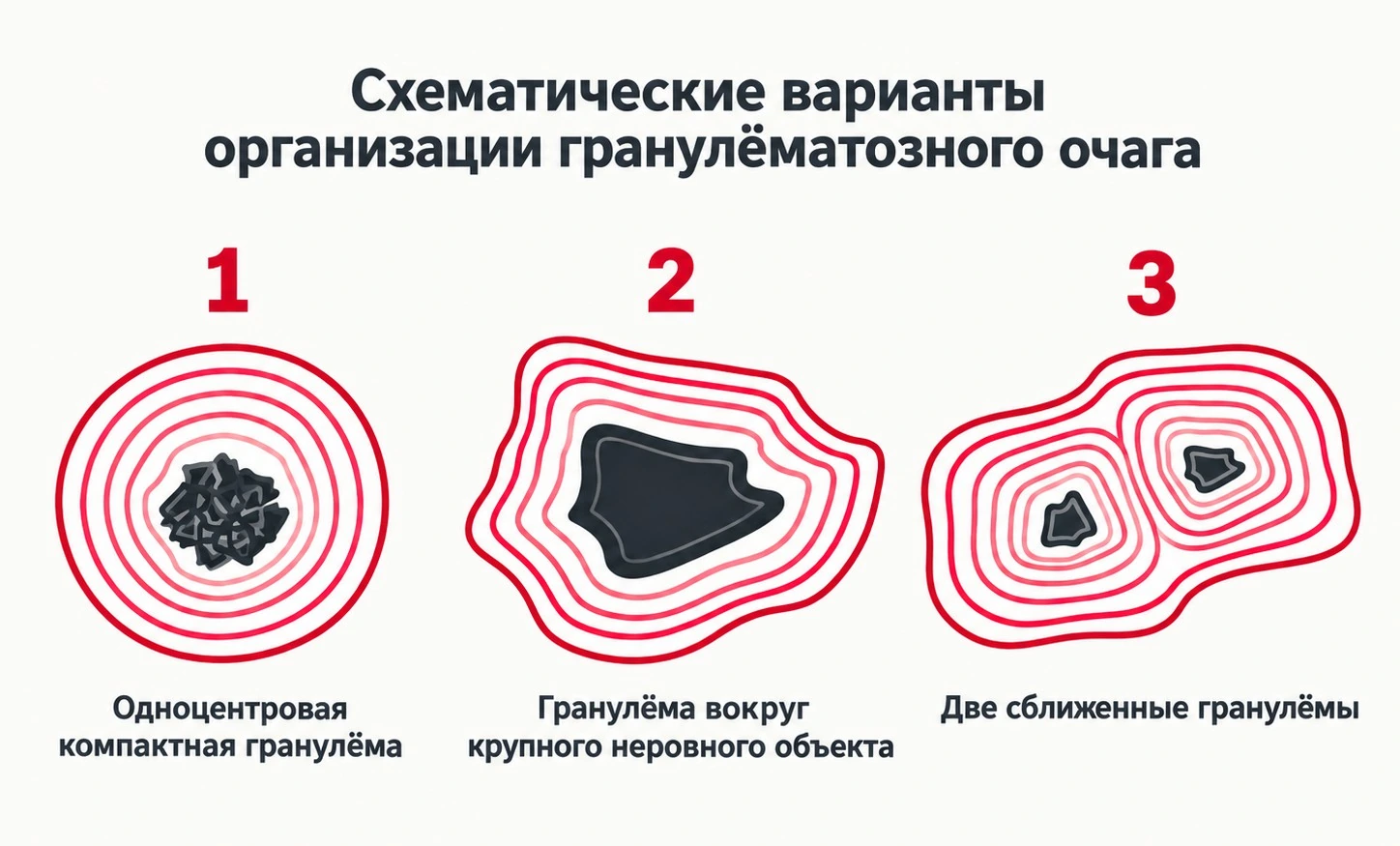
- A single compact center If the initiating factor is small, the response may organize around one center and produce a relatively round, compact granuloma.
- A large or irregularly shaped center If the underlying structure is large, uneven, or difficult to remove—such as a parasitic structure, a concretion, or a substantial area of tissue breakdown—the resulting granuloma may follow its contours and become less regular in shape.
- Two adjacent centers Sometimes two lesions lie close together in the tissue. Their outer zones of response may merge, creating the impression of a single complex granuloma. But two distinct centers suggest two closely spaced granulomas rather than one irregular lesion.
Necrotizing and non-necrotizing granulomas
Fibrous nodular lesions in the kidneys of a gudgeon (Gobio gobio). The lesions range from 0.5 to 8 mm in size. Their central regions have a homogeneous appearance. No acid-fast bacteria were detected.
Another important feature is whether necrosis is present at the center.
When cells in the center of a granuloma break down and form an amorphous mass, the lesion is termed a necrotizing granuloma.
Central necrosis is characteristic of many mycobacterial granulomas, but it also occurs in other diseases. Conversely, mycobacteriosis can occur without necrosis.
In some fungal and parasitic diseases, numerous well-organized granulomas without central necrosis may be present even in severe disseminated disease.
Within a single epizootic episode, granulomas often have similar structures. When the same agent affects a relatively uniform group of fish, the granulomatous response—its shape, capsule development, and the presence or absence of necrosis—may recur in a fairly consistent pattern. Yet this morphotype (a set of characteristic structural and shape-related features) reflects the interaction between a particular pathogen and a particular host; it is not a universal property of the pathogen.
What surrounds a granuloma?
It is important to examine not only the center of a granuloma but also its periphery. In some cases, the boundary with the surrounding tissue is poorly defined. In others, the lesion is surrounded by several layers of macrophages, other inflammatory cells, and connective-tissue elements.
Over time, a dense connective-tissue capsule may form around a granuloma. Its thickness and prominence depend on the duration of the process, the nature of the irritant, and the tissue response of the host.
Where does “tradition instead of science” come into this?
Not in the association between granulomas and mycobacteriosis itself. That association is real and can be very useful in practice. The problem arises when a probable cause is treated as the only possible cause simply because we are accustomed to seeing the two together.
The shortcut “saw a granuloma → wrote ‘tuberculosis’” saves time.
But it discards the most interesting part of diagnosis. What initiated the granuloma? Are bacteria present within it? Are they acid-fast? Is an infectious agent present at all? What is happening in the surrounding tissue? Is the process localized or generalized? Only after asking these questions do we have grounds to move from a morphological feature toward a diagnosis.
A granuloma is not a diagnosis.
It is a trace of the organism’s response.
And the task of diagnosis is to find out why that response was needed.
References
Pagán, A.J., Ramakrishnan, L., 2018. The Formation and Function of Granulomas. Annual Review of Immunology 36, 639–665.
https://doi.org/10.1146/annurev-immunol-032712-100022
A review of granuloma formation and function, including the definition of a granuloma, its evolutionary origins, infectious and noninfectious causes, responses to foreign material and persistent endogenous structures, and both the protective and pathological roles of granulomatous inflammation.
Pagán, A.J., Ramakrishnan, L., 2015. Immunity and Immunopathology in the Tuberculous Granuloma. Cold Spring Harbor Perspectives in Medicine 5, a018499.
https://doi.org/10.1101/cshperspect.a018499
A review of the interaction between mycobacteria and granulomas. Models including Mycobacterium marinum infection in zebrafish demonstrate that granulomas do not merely contain infection: mycobacteria can exploit macrophage recruitment and death to multiply and spread.
See also
Guide.
 Scientific Method. A Practical Guide.How knowledge is formed, why confidence often outpaces understanding, and how the scientific method helps distinguish observation from …
What Is Your Evidence?
Scientific Method. A Practical Guide.How knowledge is formed, why confidence often outpaces understanding, and how the scientific method helps distinguish observation from …
What Is Your Evidence?
 What Is Your Evidence?Why photographs, videos, and polished explanations are no longer enough in the age of generative AI. A practical look at provenance, …
Tradition Instead of Science
What Is Your Evidence?Why photographs, videos, and polished explanations are no longer enough in the age of generative AI. A practical look at provenance, …
Tradition Instead of ScienceI. The Method.
 Tradition Instead of Science I. The Method.What can staining still reveal when the classic Ziehl–Neelsen method cannot be performed in full? Exploring the roles of phenol, heat, and …
Initial Diagnostics After Fish
Tradition Instead of Science I. The Method.What can staining still reveal when the classic Ziehl–Neelsen method cannot be performed in full? Exploring the roles of phenol, heat, and …
Initial Diagnostics After FishTransportation
 Initial Diagnostics After Fish TransportationMicroscopy, smears, staining methods, and common mistakes in early diagnostics.
Cryptococcus-like structures in fish
Initial Diagnostics After Fish TransportationMicroscopy, smears, staining methods, and common mistakes in early diagnostics.
Cryptococcus-like structures in fish
 Cryptococcus-like structures in fishRound structures with a distinct halo, morphologically resembling cryptococci. Morphological analysis and limitations of interpretation.
Gas Bubble Disease: It's
Cryptococcus-like structures in fishRound structures with a distinct halo, morphologically resembling cryptococci. Morphological analysis and limitations of interpretation.
Gas Bubble Disease: It'sNot About the Bubbles
 Gas Bubble Disease: It's Not About the BubblesGas Bubble Disease in fish is not caused by air bubbles entering the gills but by dissolved gas supersaturation. This article explains the …