If We Change the Staining Method, What Remains of the Result?
In diagnostics, it is tempting to see a protocol as a fixed sequence of mandatory steps. If one reagent is missing, the method cannot be performed—and the specimen supposedly has nothing more to tell us.
But this confuses two different questions: Can we perform the standard test? and Can we still obtain useful observations from the specimen? The answers are not always the same.
Ziehl–Neelsen staining, used to detect acid-fast bacteria, including mycobacteria, is a good example. Conventional versions use carbol fuchsin, heating, acid or acid-alcohol decolorization, and a counterstain. Each step has its own function.
If we change a step, we need to understand what information we have preserved and what we have lost.
What Happens During Staining?
Fuchsin stains structures in a specimen. In the conventional method, phenol and heat help the dye penetrate structures, including bacteria that are difficult to stain by ordinary methods.
Then comes a fundamentally different step: acid treatment. It removes the dye from some structures, while others continue to retain it under the specified conditions. This is how we demonstrate acid-fastness—the ability to retain a stain after a particular decolorization procedure.
A counterstain helps distinguish tissues and the background.
So omitting phenol and omitting acid are not equivalent changes. In the first case, we alter the conditions of primary staining. In the second, we remove the actual test for acid-fastness.
Heating: 40 °C, 60 °C, or All the Way to Boiling?
In the conventional Ziehl–Neelsen method, carbol fuchsin is heated after it has been applied to the specimen, usually until vapors begin to appear. Some protocols associate this visual cue with approximately 60 °C. The dye is kept heated for several minutes (often 3–5 minutes), without allowing it to boil or dry out ( ASM protocol; comparative study).
But the appearance of vapors is not the same as boiling: liquids evaporate at temperatures below their boiling points. Thus, “until vapors appear” is only an approximate guide, not a precise way to control temperature. For a reproducible modified procedure, it is better to record the measured dye temperature, exposure time, and concentration.
Why not heat it further? Boiling is not the goal of the method. It increases evaporation and can cause the dye to dry out or splatter. With phenol, chemical safety is an additional concern. It is also important to distinguish heat fixation of the specimen before staining from heating the dye during staining—these are separate operations.
Phenol-free methods use different conditions. Some studies used 0.5% aqueous fuchsin at 60 °C for 15 minutes; the GASMoC method used 1% aqueous fuchsin at 37 °C for 15 minutes. Staining at 37–40 °C is therefore possible in principle, but 40 °C is not a universally validated temperature: any protocol has to be evaluated in terms of dye concentration, exposure time, and specimen type. These findings apply to specific studies; they do not establish that all the conditions are interchangeable.
Can We Do Without Phenol?
Yes, under certain conditions. This is supported by published research, not just isolated observations.
Della Speranza and Fail (2000) showed that a 0.5% aqueous solution of basic fuchsin, without phenol, could stain mycobacteria at 60 °C in 15 minutes. At room temperature, the same approach performed considerably worse under their conditions. So the outcome depends not only on the composition of the stain, but also on how it is applied ( 2000 study).
McCollough (2008) used staining with 0.5% aqueous fuchsin at 60 °C for 15 minutes to examine pathogens of aquatic animals, including Mycobacterium marinum in the kidney of a goldfish ( study).
Gomes et al. (2020) compared conventional staining with a modified phenol-free procedure using 1% aqueous fuchsin at 37 °C for 15 minutes, both on clinical samples and on zebrafish tissue containing M. marinum. In the samples studied, the new procedure was not inferior to the conventional method for detecting acid-fast bacteria. However, the sample size was small, and the findings cannot simply be generalized to all specimens and conditions ( GASMoC study).
One point matters in all these cases: acid decolorization was retained. The researchers changed the primary stain, but they did not abandon the test for the property they were investigating.
Therefore, the absence of phenol does not, by itself, make it impossible to detect acid-fast bacteria. But the conditions of a particular staining procedure must be evaluated, and its results need appropriate controls.
What Happens If We Leave Out the Acid?
Suppose that, after intensive fuchsin staining, red rod-shaped bacteria are visible in the tissue. We can examine their shape, approximate numbers, distribution, and association with altered regions of the organ.
But if acid decolorization was not performed, acid-fastness was not tested. We cannot report “acid-fast bacteria detected,” however convincing the rods may look.
There is another side to this change. Acid can remove stain not only from the background but also from structures we might want to examine. Without acid, the microscopic field may look more cluttered, but additional objects may remain visible. This can be useful during exploratory examination, although it reduces the selectivity of the procedure.
Therefore, staining without acid decolorization is not a simplified Ziehl–Neelsen test. It is a different procedure with a different set of permissible conclusions.
One Organ, Three Methods
For my own comparison, I used specimens prepared from the same angelfish spleen, which contained numerous pale lesions. The specimens were stained in three ways:
- With phenol, followed by acid decolorization.
- Without phenol, but with decolorization using 5% sulfuric acid.
- Without phenol and without acid decolorization.
All specimens were imaged using the same microscope and camera.
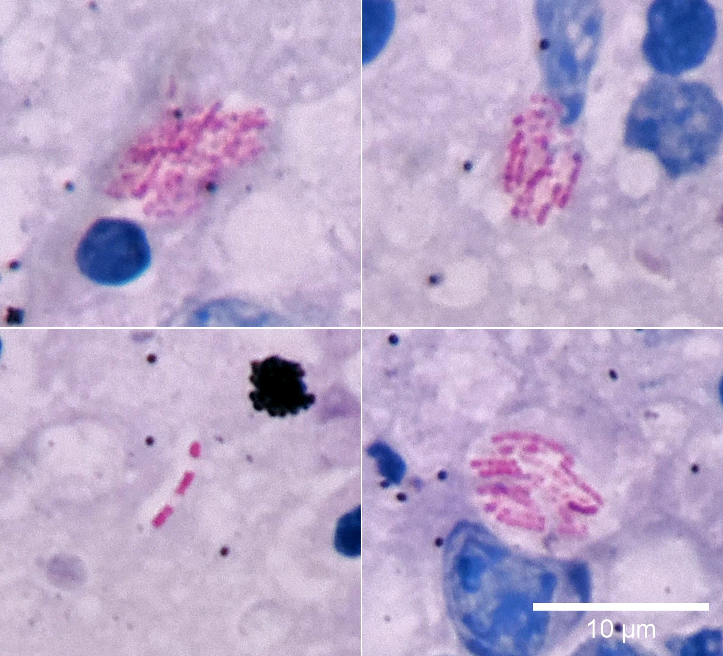
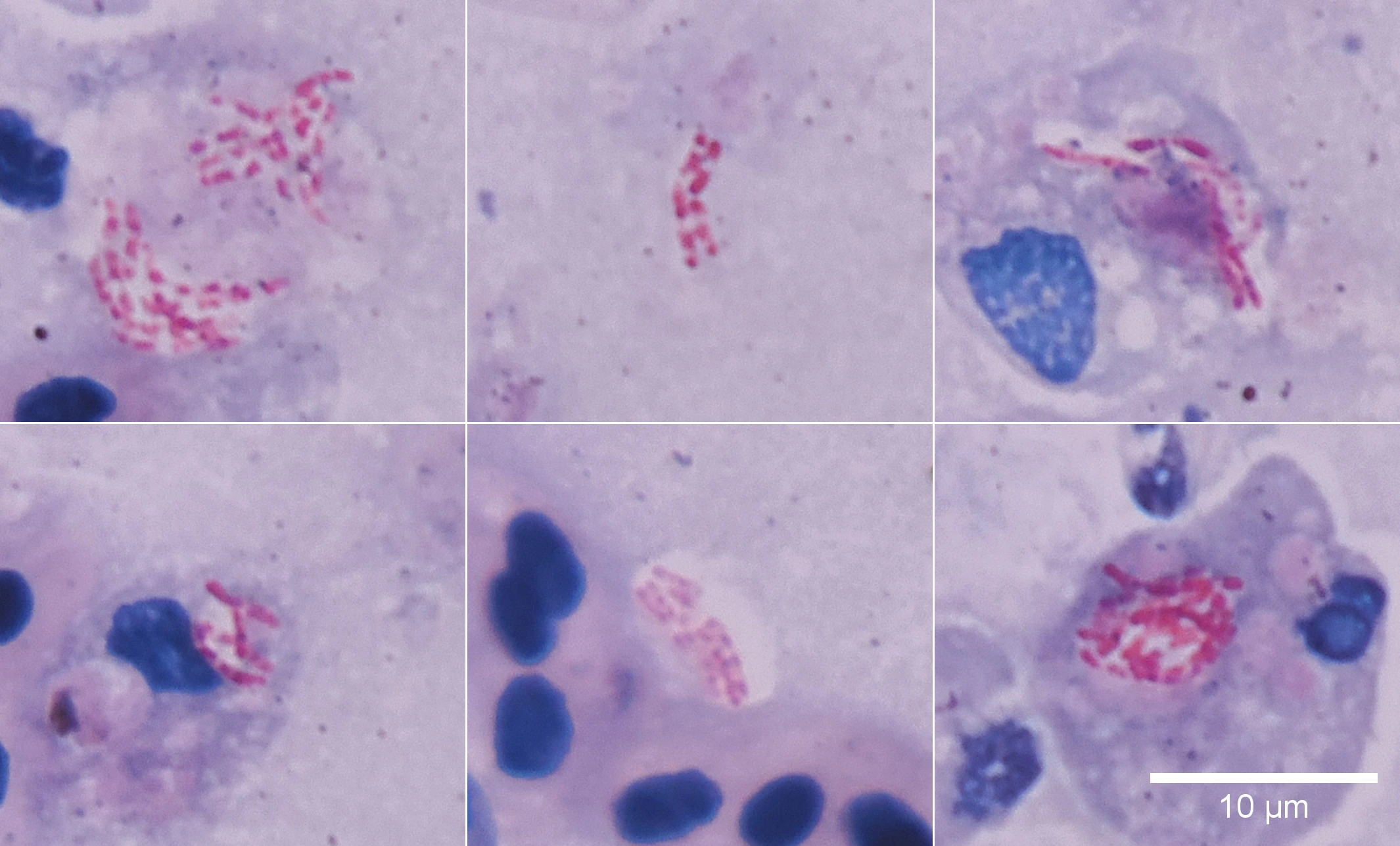
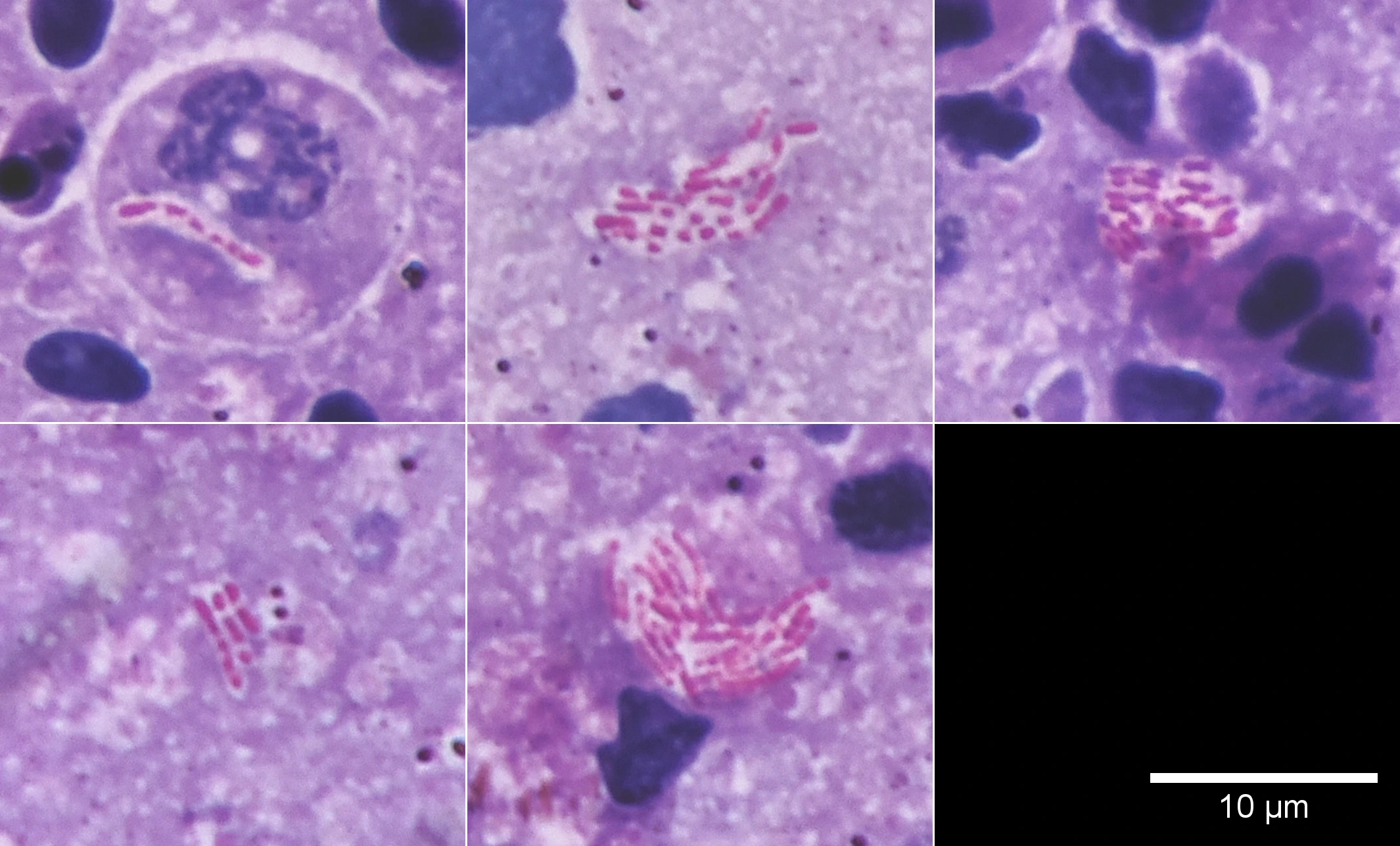
Similar red rod-shaped bacteria were observed in all three preparations. This is consistent with the possibility of staining without phenol, including retention of the stain after acid treatment.
However, specimens from a single organ do not allow us to calculate sensitivity or specificity or demonstrate that the three procedures are equivalent. This is a comparison of observed results, not validation of a new diagnostic method.
What If We Use a Different Acid?
Different acids and decolorization conditions have been used in historical and modern acid-fast staining methods. Thus, no single acid is a universal, indispensable component of every variant of the method ( Vilchèze and Kremer, 2017).
But this does not mean that any acidic solution provides the same test. The type and concentration of the acid, as well as the duration of treatment, affect which structures lose their stain.
For example, a concentrated citric acid solution could be considered an experimental decolorizing agent. Even if the background loses its color while the rods remain red, this is not automatically equivalent to a standard test. Appropriate positive and negative control specimens would be needed for comparison.
A Negative Staining Result Has Limits, Too
There is another reason not to turn a staining result into a definitive diagnosis.
Even mycobacteria do not always exhibit acid-fastness to the same degree. In Mycobacterium tuberculosis, states have been described in which some cells are poorly detected or not detected at all by conventional staining. This is discussed in the work on the so-called “Koch’s paradox” ( Vilchèze and Kremer, 2017).
For fish diagnostics, the cautious principle is this: a negative staining result does not, by itself, prove that mycobacteria are absent. But neither can every negative stain of fish tissue be explained by “dormant” mycobacteria. That is only one possible explanation and would require investigation.
Conversely, a positive acid-fast staining result does not automatically establish the genus or species of the bacteria. Additional testing, such as PCR or culture, may be needed for identification.
How to Describe the Finding Correctly
If heated fuchsin was used without subsequent acid decolorization, the report should describe both the procedure and what was actually observed:
A modified fuchsin staining procedure with heating, but without acid decolorization, was used. Intensely stained rod-shaped bacteria were observed in the tissue. Their acid-fastness was not assessed.
It would be incorrect to state that standard Ziehl–Neelsen staining had been performed and that the presence of acid-fast bacteria had been demonstrated.
The key distinction: omitting a step does not invalidate the observations already made, but it changes the limits of the conclusions we can draw. A method is not a magic formula that must be recited word for word; it is a sequence of actions, each of which should answer a specific question.
For the history of these modifications, the exact experimental conditions, and a detailed bibliography, see the full F.O.S. version. The F. version tells the same story with almost none of the laboratory details.
See also
Late for a Fish
Autopsy?
 Is It Ever Too Late for a Fish Autopsy?Debunking the myth: why you can examine a fish hours after death—not just in the first 30 minutes.
What Is Your Evidence?
Is It Ever Too Late for a Fish Autopsy?Debunking the myth: why you can examine a fish hours after death—not just in the first 30 minutes.
What Is Your Evidence?
 What Is Your Evidence?Why photographs, videos, and polished explanations are no longer enough in the age of generative AI. A practical look at provenance, …
Tradition Instead of Science
What Is Your Evidence?Why photographs, videos, and polished explanations are no longer enough in the age of generative AI. A practical look at provenance, …
Tradition Instead of ScienceI. The Method.
 Tradition Instead of Science I. The Method.What can staining still reveal when the classic Ziehl–Neelsen method cannot be performed in full? Exploring the roles of phenol, heat, and …
Cryptocaryonosis: killing the parasite
Tradition Instead of Science I. The Method.What can staining still reveal when the classic Ziehl–Neelsen method cannot be performed in full? Exploring the roles of phenol, heat, and …
Cryptocaryonosis: killing the parasiteor treating the water?
 Cryptocaryonosis: killing the parasite or treating the water?A practical analysis of cryptocaryonosis treatment in marine fish: why killing the parasite should not become more important than the …
Fish coccidia: Goussia
Cryptocaryonosis: killing the parasite or treating the water?A practical analysis of cryptocaryonosis treatment in marine fish: why killing the parasite should not become more important than the …
Fish coccidia: Goussia
 Fish coccidia: GoussiaCoccidia of the genus Goussia in fish. Morphology of oocysts, sporocysts, and sporozoites, with diagnostic interpretation of microscopic …
Two Sides of One
Fish coccidia: GoussiaCoccidia of the genus Goussia in fish. Morphology of oocysts, sporocysts, and sporozoites, with diagnostic interpretation of microscopic …
Two Sides of OneDosage
 Two Sides of One DosageAquarium medications: remedy or threat? Pros and cons of antibiotics, formalin, methylene blue, malachite green, and the FMC mixture.