Microscopic Observations
Videos, time-lapses and microscopic observations
-
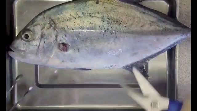
Fish necropsy Fish necropsy with examination of internal organs and pathological findings. -
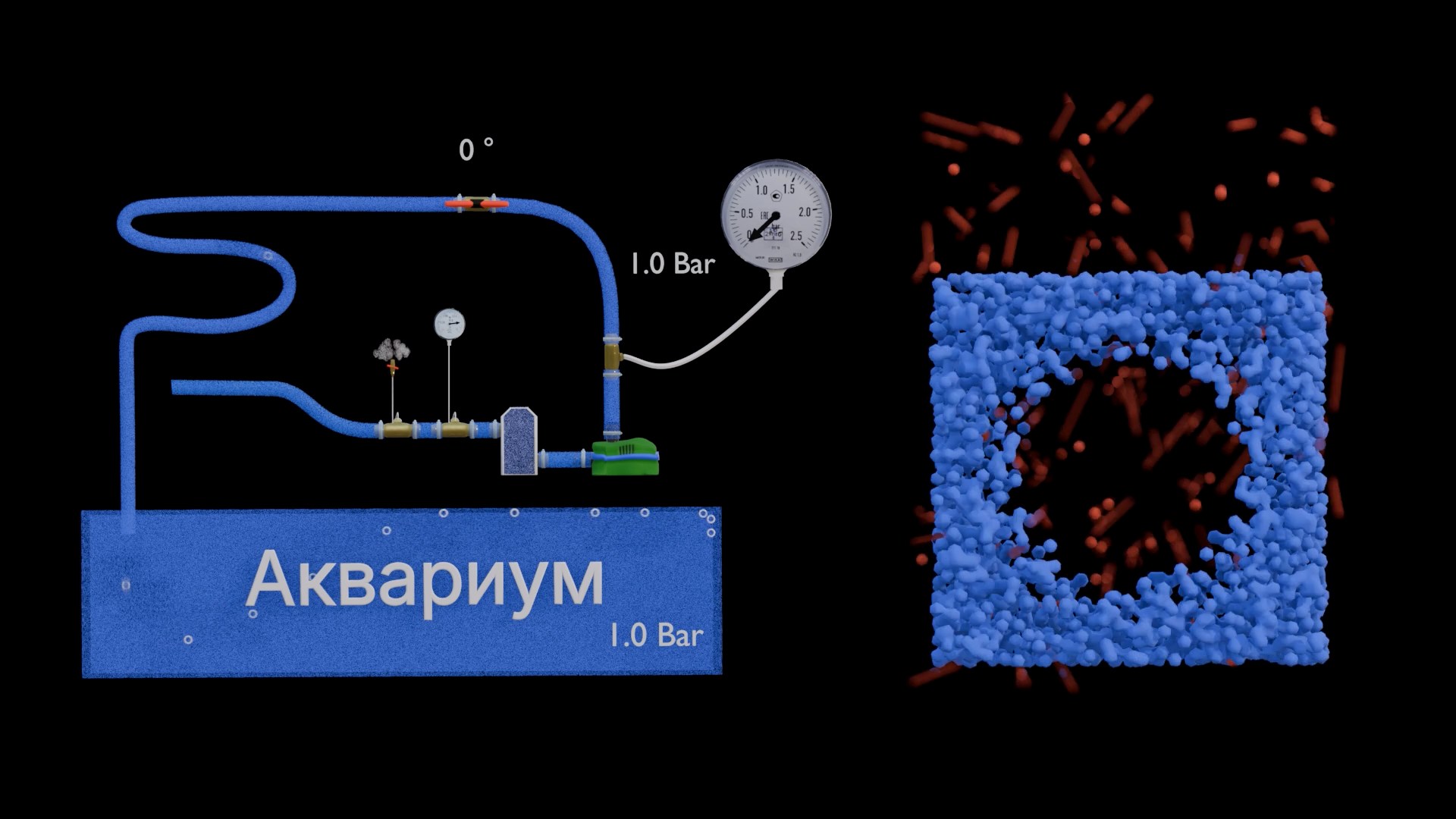
Bubble formation 3D visualization of bubble formation in supersaturated water. -
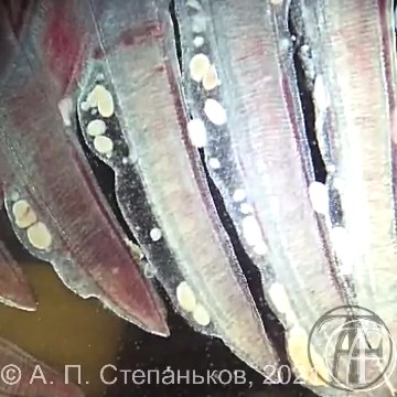
Cryptocaryon irritans in gills Cryptocaryon irritans trophonts within the gill epithelium. The video demonstrates the gill-dominant form of cryptocaryoniasis, which may occur without the typical white spots on the skin. -
Fin ray structure A fin ray consists of two paired halves separated by living tissue. This video demonstrates fin ray anatomy and helps explain the mechanisms of fin erosion. -
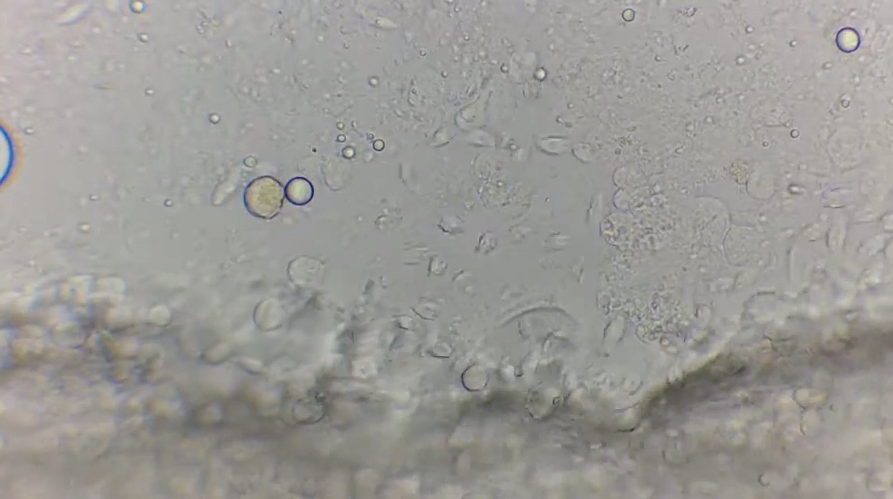
What 'many hexamitids' actually looks like Dense aggregation of flagellated protozoans in a wet mount. The video demonstrates a parasite burden that is difficult to appreciate from still images alone. -
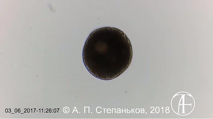
Division of Ichthyophthirius multifiliis Palintomic division of a tomont after leaving the fish. -
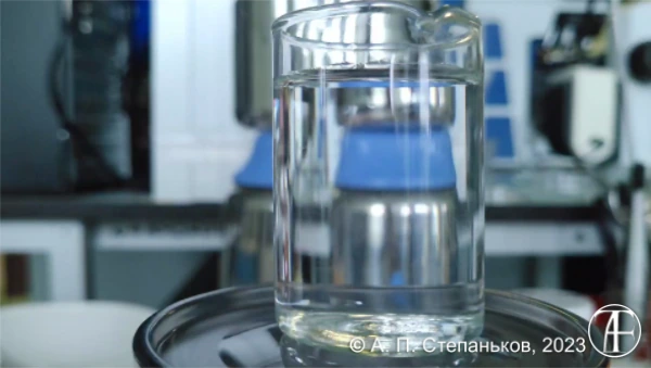
Praziquantel in Water Behavior of praziquantel after addition to water. Despite prior dissolution in alcohol, the compound forms a fine suspension and gradually settles. The video illustrates why the term "dispersion" is often more appropriate than "dissolution" in aquarium treatments. -
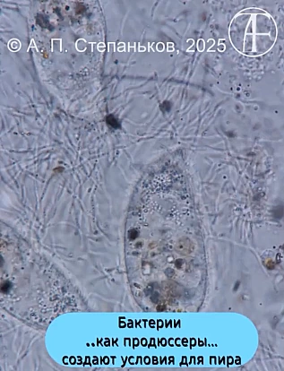
Ciliates and Bacteria on Fish Surfaces The video shows free-living ciliates found on the surface of a fish together with bacterial aggregates among which they occur. Attention is often focused on the conspicuous and motile ciliates, whereas the bacterial component may remain unnoticed. Both native and stained preparations are presented to illustrate the spatial relationship between ciliates and bacteria. The footage demonstrates that the presence of ciliates alone does not explain the cause of a lesion and should be interpreted together with tissue condition and associated microbial communities. -
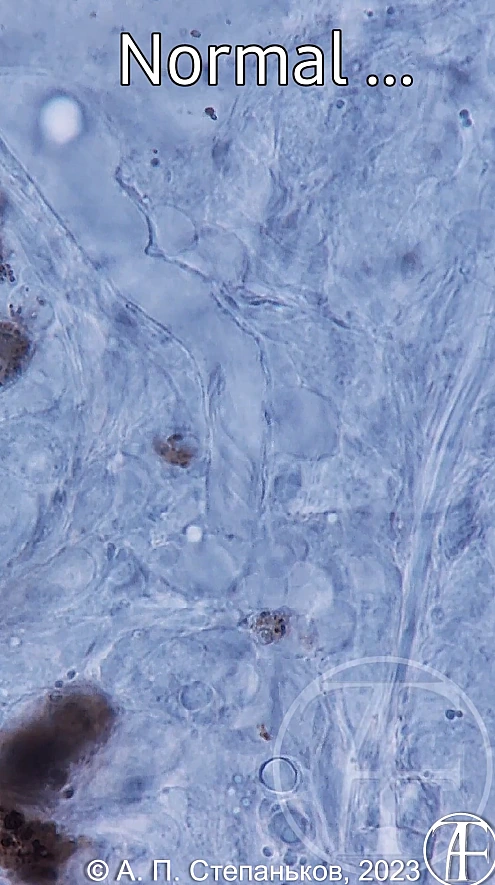
Ciliated Epithelium of the Kidney Ciliated epithelium within the renal tubules of a fish. The video shows ciliary motion in real time and then in slow motion, allowing the beating pattern to be observed in greater detail. -
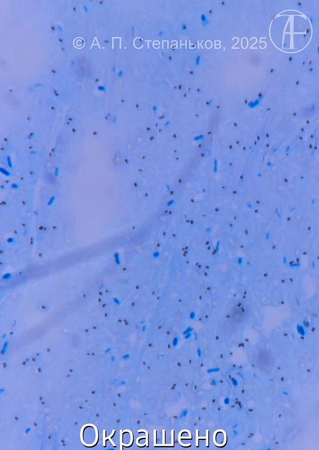
Pigment or Cocci? Not every dark particle in a smear is a coccus. The same microscopic field is shown after staining and subsequent alcohol decolorization, illustrating the difference between bacterial structures and pigment granules. -
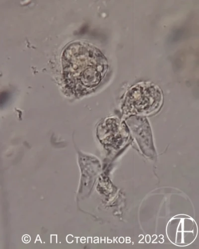
Cryptobia Moving Through Detritus The video shows flagellates of the genus Cryptobia actively moving between particles of detritus and mucus. Their characteristic locomotion, frequent changes in direction, and ability to pass through narrow spaces are clearly visible. Internal structures, including food vacuoles, can be observed in some individuals. The footage illustrates how these protozoans navigate through their natural microenvironment. -
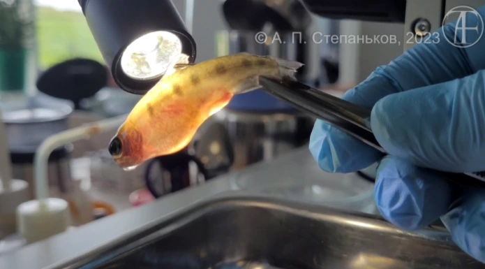
Fungal Hyphae in Fish Tissues Free spores alone do not prove tissue invasion. The video demonstrates the diagnostic sequence from external examination and hemorrhages to the detection of fungal hyphae actively growing within fish tissues. -
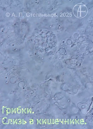
Fungal Structures in a Native Preparation Native preparation of fungal structures within fish tissue. The video shows hyphae, spores, and their arrangement without fixation, drying, or staining. This approach preserves the natural size and morphology of fungal elements, facilitating their recognition and comparison with stained preparations. The footage complements diagnostic materials on fungal lesions and demonstrates the appearance of spores and hyphae before artefacts associated with smear preparation are introduced. -
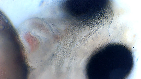
Heart of an Amur Sleeper Larva Under the Microscope The video shows the functioning heart of a rotan larva (Perccottus glenii). Due to the transparency of larval tissues, contractions of the atrium and ventricle, blood flow, and the operation of a two-chambered teleost heart can be observed directly. The footage illustrates basic aspects of fish circulation and provides a rare opportunity to observe cardiac activity without dissection or staining. -
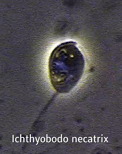
Ichthyobodo spp.: Comparing Morphotypes The video compares two morphotypes of flagellates traditionally identified as Ichthyobodo spp. Differences in cell morphology and flagellar arrangement become apparent at high magnification. However, the available morphological features are insufficient for reliable species identification, and the taxonomic status of these forms remains uncertain. -
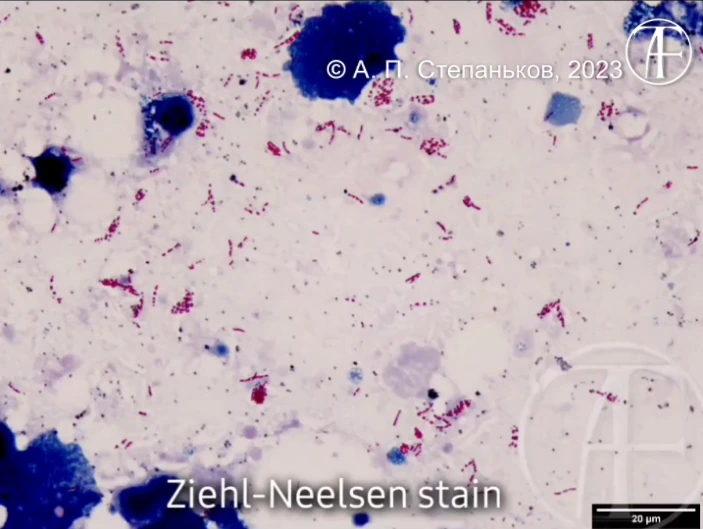
Loeffler vs Ziehl–Neelsen The same smear is shown sequentially after Loeffler’s methylene blue stain and Ziehl–Neelsen staining. The video demonstrates how acid-fast bacteria become visible following a specialized staining procedure.