Fungal elements are regularly encountered in fish preparations.
However, their presence alone does not allow a definitive conclusion about the nature of the process. In some cases, this represents surface colonization of damaged tissues; in others, an invasive process with spread into tissues and body cavities.
This page presents observations of fungal structures in fish and an approach to their interpretation.
Saprolegnia and surface colonization
The most well-known “fungal” lesions in fish are associated with Saprolegnia. In aquaculture and aquariums, this appears as a characteristic “cotton-like” growth on the body. In the vast majority of cases, Saprolegnia is not the cause but a consequence of tissue damage, necrosis, or stress (transport, aggression, environmental conditions).
Under normal conditions, the fish immune system effectively suppresses these organisms, despite the constant presence of spores in the water. When defenses are compromised, the organism begins to develop actively and forms superficial mycelial masses. This is why juveniles are affected more frequently: less developed immunity, smaller body size, and less time to compensate for damage.
It is important to note that Saprolegnia is not a “true fungus” but an oomycete.
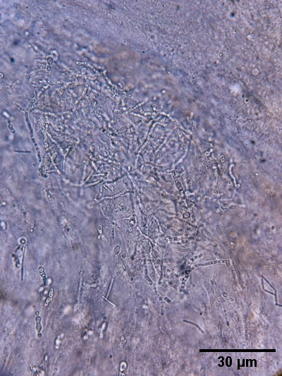
Nevertheless, morphologically it forms typical mycelial structures.
Microbial communities
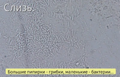
Superficial mycelial growth rarely exists in isolation. Hyphae form a structure that harbors bacteria, ciliates, nematodes. In practice, this represents a complex microbial system rather than a purely fungal process.
Parasitic forms
There are cases where fungal elements behave differently.
Unlike Saprolegnia, hyphae do not form prominent tufts but spread along tissue surfaces, intertwine, and form dense слизистые structures. Such processes may be associated with involvement of internal organs.
Observation
Below is an example observed in juvenile salmonids.
Typical findings:
- gills pale, light pink,
- internal organs pale, covered with transparent mucus,
- stomach and intestine filled with mucus,
- fungal spores present in contents and on organ surfaces.
Such conditions are often accompanied by intestinal distension.
Important:
bacterial gas production is typically postmortem,
whereas similar changes in fungal processes may develop during life.
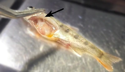
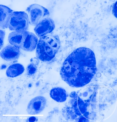
Organs within the body cavity are covered with mucus.
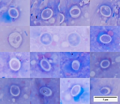
Spores are readily detected in the mucous content of internal organs.
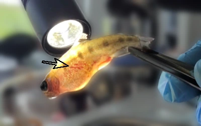
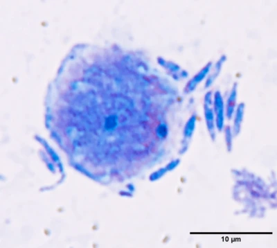
On transmitted light, hemorrhages can be observed in the abdominal region.
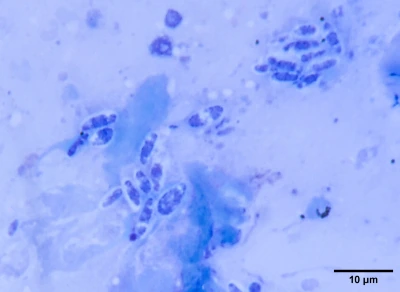
Hyphae and structures associated with sporulation are detected in scrapings.
The picture becomes coherent:
- hyphae are localized in tissues and cavities,
- spores are present in mucus,
- changes are systemic.
This allows morphological findings to be interpreted as a unified process.
Staining may result in a reduction in spore size, which can complicate interpretation.
Therefore, for primary detection, native preparations and water-based stains (e.g., Löffler) are preferable.
Fungal structures in fish may represent either secondary colonization or an independent pathological process.
Their evaluation should be based not on the fact of detection,
but on morphology, localization, and context.