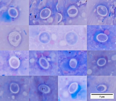
Two Spleens. One Diagnosis?
Here are two photographs of fish spleens. Both have numerous whitish spots and nodules. At first glance, they look similar.
Someone familiar with fish diseases might suspect mycobacteriosis. Or, out of habit, use an even simpler term: fish tuberculosis.
There is a reason for that suspicion. Mycobacterial infections often cause nodular lesions in fish organs. But similar appearances do not necessarily mean the same cause.
Let’s see what was found in these two cases.
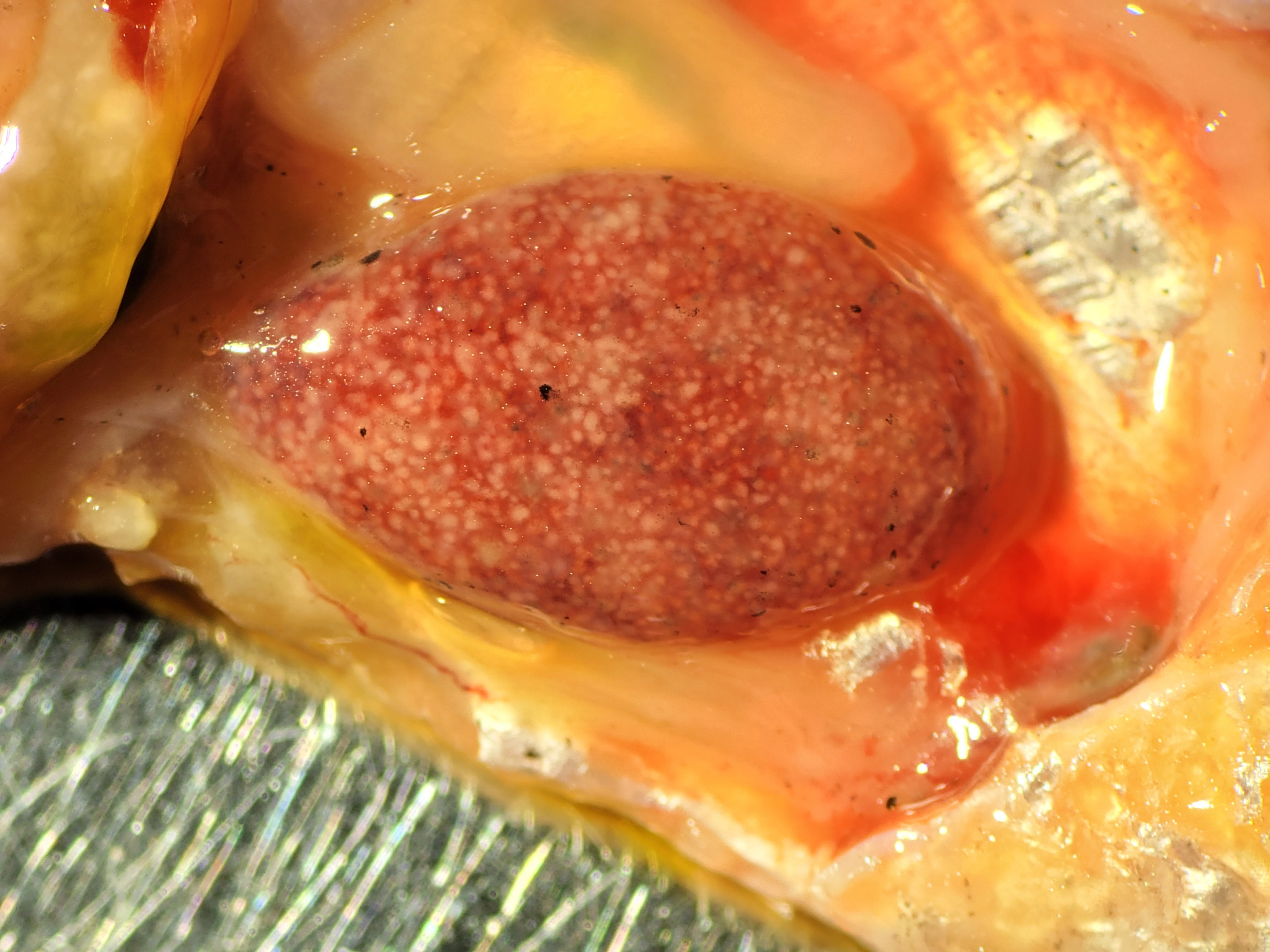
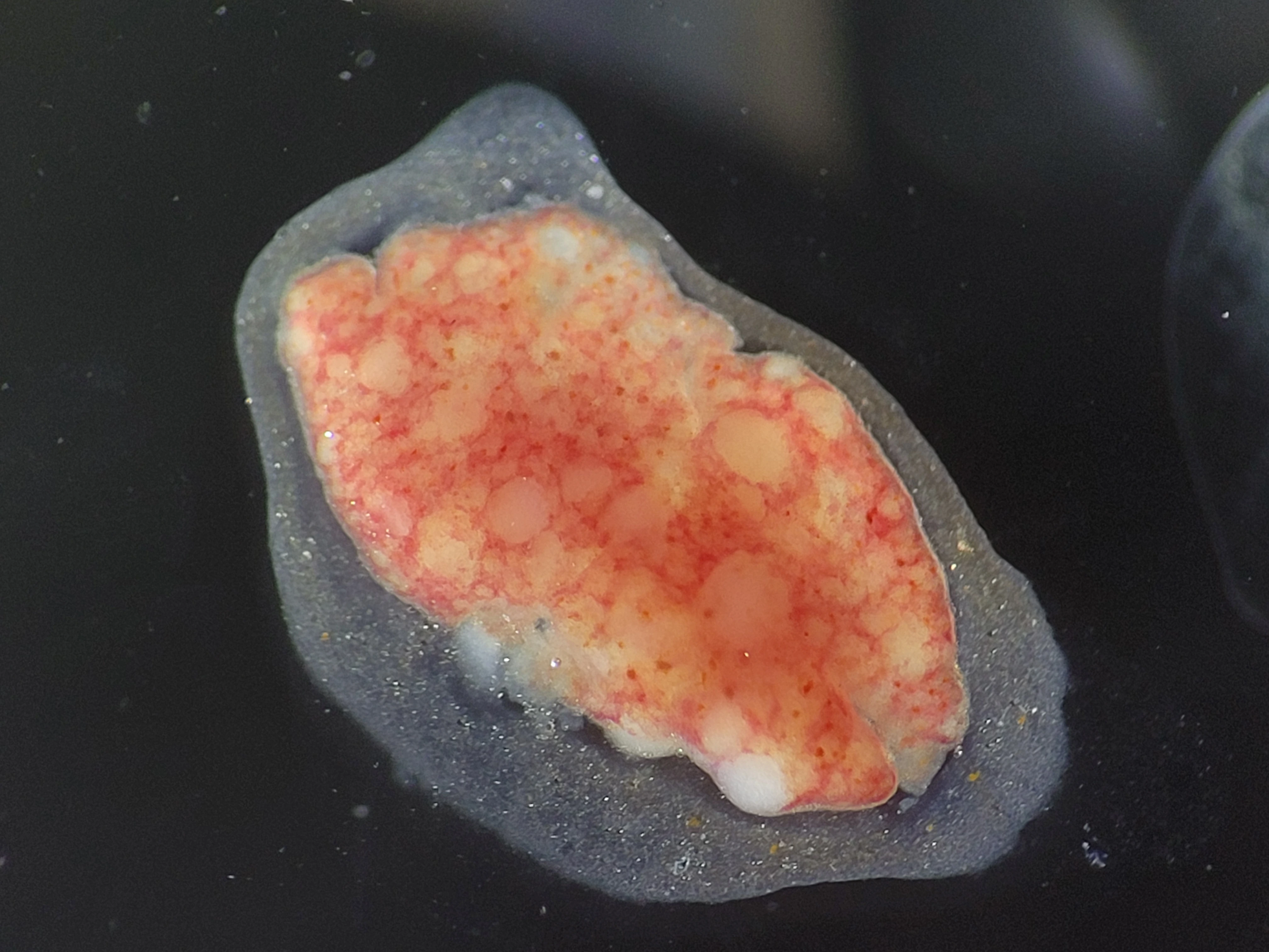
And there is the surprise: they look alike from the outside, but the microscopic findings are different. Seeing only the white spots, we could not have known in advance what we would find inside.
So What Is a Granuloma?
Sometimes, a necropsy reveals pale nodules. When the tissue is examined under a microscope, some of these turn out to be organized clusters of immune cells. These are called granulomas.
One job of these cells is to engulf and remove things that are causing trouble. They can take up bacteria, the remains of dead cells, and other material. But not everything can be broken down and cleared away quickly.
When the problem persists, immune cells may gather around it and form an organized focus. This is one way a granuloma can develop.
An important distinction: not every white nodule seen at necropsy is necessarily a granuloma. To call something a granuloma, we need to examine its structure, not just its color and outward shape.
Why a Granuloma Does Not Identify a Single Culprit
Imagine finding such a cluster of cells in a tissue. What does it tell us?
Perhaps the body is trying to contain bacteria. But the cause might also be another microorganism, a parasite, a foreign particle, or material left behind by damaged tissue that the body cannot easily remove.
In other words, a granuloma shows how the body is responding to a problem, but it does not tell us what that problem is.
Think of it as finding a repair crew at work. The workers tell us that something has happened, but without inspecting the site, we cannot know whether the problem is a broken pipe, faulty wiring, or a damaged wall.
Granulomas have another twist: in certain infections, a cluster of immune cells may not only contain the pathogen but also help it spread. That is why the number and appearance of granulomas cannot be used as a simple count of the bacteria present.
What If We Find Red Rods?
One way to investigate suspected mycobacteriosis is to stain a tissue preparation. After treatment with a dye and an acid, some bacteria remain red. This property is called acid-fastness.
If rod-shaped bacteria with these staining properties are found in the affected tissue, the suspicion of mycobacteriosis becomes stronger. But their color alone does not tell us which bacterial species they belong to. Further tests may be needed.
Conversely, if acid-fast bacteria are not detected, that does not mean every possible cause of the lesions has been ruled out.
We explain the staining procedure and its limitations in “Tradition Instead of Science I: Method”.
So What Should We Write in the Report?
Start with what has actually been established.
If all we can see at necropsy are whitish nodules, we can describe them exactly that way. If microscopy confirms that they are granulomas, we can write: “multiple granulomas.”
But a diagnosis of mycobacteriosis requires evidence of its own.
Similar shape, white color, necrosis, or a large number of nodules cannot replace an investigation into their cause.
A granuloma is not a diagnosis. It is a trace of the body’s response. The task of diagnosis is to find out what made that response necessary.
Want to explore the topic further? F.O. — how granulomas form and what morphology can tell us · F.O.S. — a full analysis with microscopic preparations and references.
See also
 Flying ElephantsWhy science requires more than an elegant hypothesis. The role of photographs, videos, specimens, and primary data in building and verifying …
Tradition Instead of Science
Flying ElephantsWhy science requires more than an elegant hypothesis. The role of photographs, videos, specimens, and primary data in building and verifying …
Tradition Instead of ScienceI. The Method.
 Tradition Instead of Science I. The Method.What can staining still reveal when the classic Ziehl–Neelsen method cannot be performed in full? Exploring the roles of phenol, heat, and …
Traditions Instead of Science.
Tradition Instead of Science I. The Method.What can staining still reveal when the classic Ziehl–Neelsen method cannot be performed in full? Exploring the roles of phenol, heat, and …
Traditions Instead of Science.II
 Traditions Instead of Science. IIWhy a familiar treatment protocol does not automatically become evidence-based therapy. Using FMC as an example, we examine how …
Cryptocaryonosis: killing the parasite
Traditions Instead of Science. IIWhy a familiar treatment protocol does not automatically become evidence-based therapy. Using FMC as an example, we examine how …
Cryptocaryonosis: killing the parasiteor treating the water?
 Cryptocaryonosis: killing the parasite or treating the water?A practical analysis of cryptocaryonosis treatment in marine fish: why killing the parasite should not become more important than the …
Granulomas and intracellular agents
Cryptocaryonosis: killing the parasite or treating the water?A practical analysis of cryptocaryonosis treatment in marine fish: why killing the parasite should not become more important than the …
Granulomas and intracellular agentsin fish
 Granulomas and intracellular agents in fishA collection of real observations: intracellular yeast-like organisms, coccidia, cryptosporidia, and fungal structures in fish.
Hexamitids Without Myths
Granulomas and intracellular agents in fishA collection of real observations: intracellular yeast-like organisms, coccidia, cryptosporidia, and fungal structures in fish.
Hexamitids Without Myths
 Hexamitids Without MythsWhy hexamitids are not a normal component of fish intestinal microflora, where the myth of 'internal parasites' comes from, and how …