When Red Rods Matter More Than the Name of the Method
Imagine examining tissue from a sick fish under a microscope. You can see cells, patches of material, and fragments of tissue. But are there any bacteria among them? It’s hard to tell.
Laboratories use special staining methods for situations like this. A stain makes structures visible that would be difficult to distinguish in an unstained specimen.
In this case, we use fuchsin—a dye that stains certain cells and bacteria bright red. It lets us see structures under the microscope that would otherwise be difficult to make out. In the conventional method, phenol is added to the fuchsin. Phenol is a chemical that does not stain anything by itself, but helps the dye penetrate bacterial structures that are difficult to stain. This mixture is called carbol fuchsin.
Heating also matters. In conventional Ziehl–Neelsen staining, the dye is gently heated until faint vapors appear—approximately 60 °C. It does not need to boil. Heat helps fuchsin stain bacteria, but that does not mean hotter is always better.
After staining, the specimen is treated with acid. The acid removes the dye from many structures, while certain bacteria remain red. This is how we test for acid-fastness—the ability to retain the stain after acid treatment.
But the necessary reagents are not always available. So what should we do—put the specimen aside and accept that we cannot learn anything from it?
I’d rather ask a different question: what can we see if we change the usual staining procedure? And, just as importantly, what can we legitimately conclude from the result?
One Angelfish, Three Specimens
Let’s take the spleen of an angelfish with numerous pale spots. We prepare specimens from this tissue and stain them in three different ways.
In the first, we follow the conventional procedure: a stain containing phenol, followed by acid treatment. In the second, we leave out the phenol but keep the acid treatment. In the third, we use neither phenol nor acid decolorization.
Why do this? Not to give an old method a new name. We want to see how much each step actually matters.
The results are easy to see. All three preparations contain clearly visible small red rods, similar in shape and distribution.
We use the same microscope and camera for comparison.
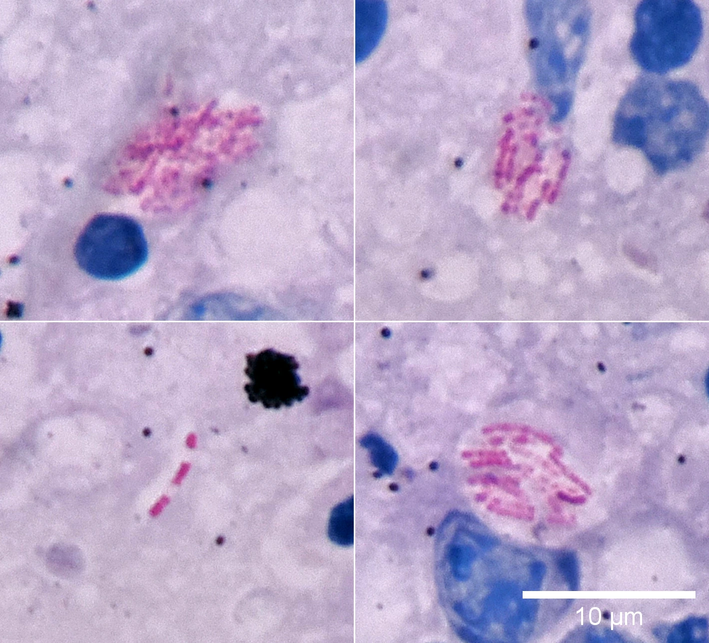
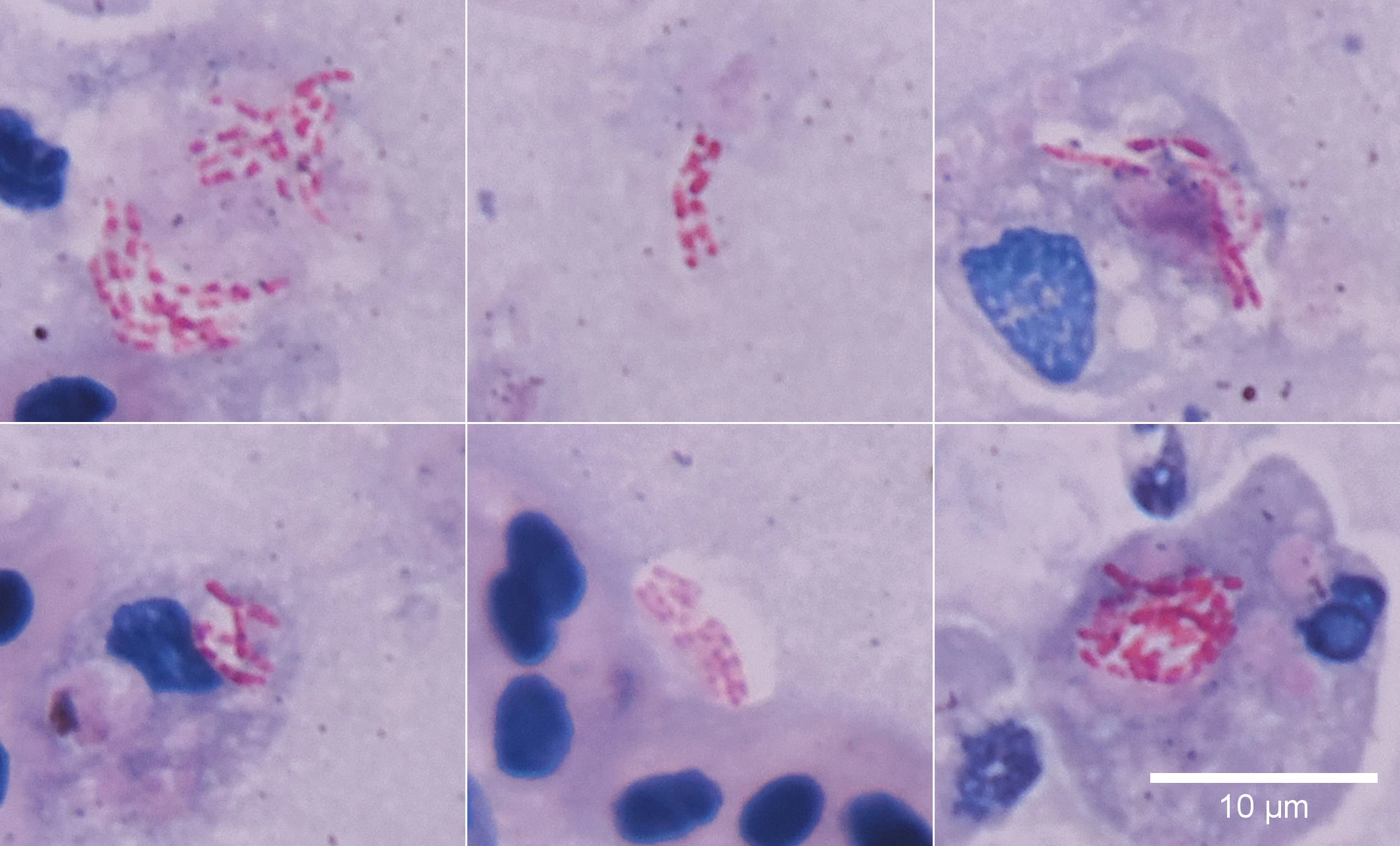
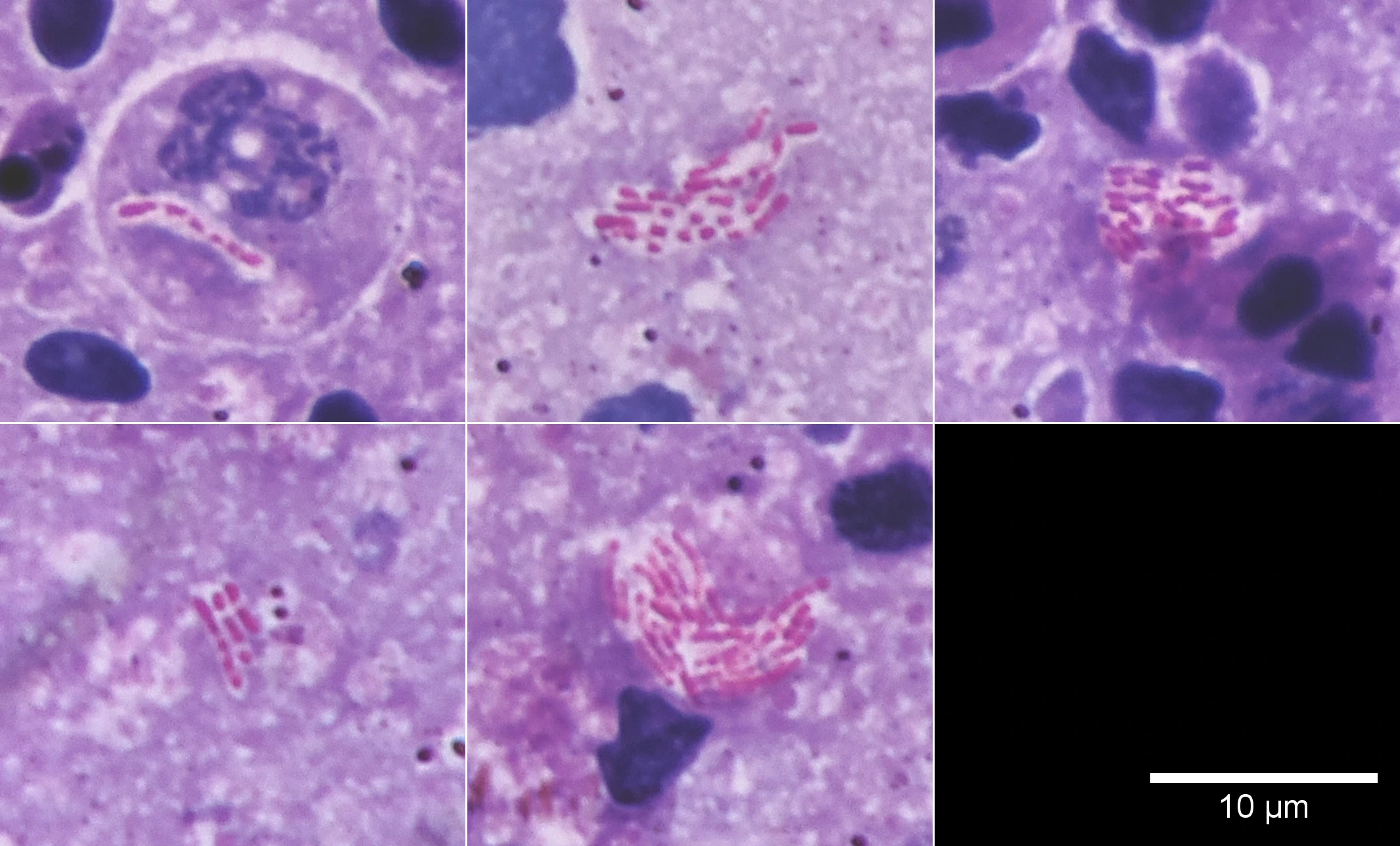
This is not a large scientific study, just a comparison of several specimens from the same organ. These images are not enough to claim that all three methods are equally reliable. But they do demonstrate one useful point: changing the procedure does not necessarily make the result disappear.
Why You Cannot Simply Skip the Acid
There is an important distinction here.
Some bacteria remain red even after acid treatment. This property is called acid-fastness. It is what the well-known Ziehl–Neelsen staining method is designed to test.
If we did not treat the specimen with acid, we did not perform that test. Even if the rods look exactly like those in another preparation, their color alone does not justify calling them acid-fast.
But does that make the rods we can see useless? Not at all. We can still examine their shape, number, distribution, and the areas of tissue in which they appear.
The distinction is simple:
Seeing bacteria is one task. Testing whether they are acid-fast is another. Identifying which bacteria they are is a third.
One photograph cannot answer all three questions at once.
So What Is the Phenol For?
Phenol is part of the conventional staining solution and helps the staining process. But “helps” does not mean “cannot be done without it.”
Researchers have already shown that, under certain conditions, bacteria can be stained with fuchsin without phenol. Temperature turned out to be particularly important. For example, aqueous fuchsin was successfully used at 60 °C for 15 minutes, including on fish tissues ( McCollough, 2008). Another study achieved staining at just 37 °C for the same 15 minutes by changing the dye concentration ( Gomes et al., 2020). Both methods retained the subsequent acid decolorization step.
So the question “40 degrees, 60 degrees, or all the way to boiling?” cannot be answered with a single number. The result also depends on the solution’s composition and the staining time. 37 °C was tested in a particular protocol; that does not prove every staining procedure will work at 40 °C. And boiling is not necessary at all.
The point, then, is not whether phenol is good or bad. The question is what function it serves, and whether that function can be achieved another way.
Why This Matters
When working in diagnostics, you may lack not only reagents but also time, equipment, or the opportunity to send a sample to a laboratory right away.
In that situation, it is easy to fall into one of two extremes. The first: “If I cannot do it exactly by the book, there is no point looking at anything.” The second: “I see red rods, so the diagnosis is confirmed.”
Both get in the way of thinking.
A microscope can provide useful information even when a definitive answer is still out of reach.
We just have to avoid claiming more than the evidence shows.
You can change a method. But then you need to ask yourself again: “What did I actually test? What did I observe? And what do I still not know?”
That is the difference between following instructions and understanding why those instructions exist.
Want to explore the subject further? Read how the staining works and where the limits of interpretation lie or the full discussion, including the method’s history, experiments, and sources.
See also
Transportation
 Initial Diagnostics After Fish TransportationMicroscopy, smears, staining methods, and common mistakes in early diagnostics.
Gas Bubble Disease. Part
Initial Diagnostics After Fish TransportationMicroscopy, smears, staining methods, and common mistakes in early diagnostics.
Gas Bubble Disease. PartII. Consequences.
 Gas Bubble Disease. Part II. Consequences.Comparison of normal and pathological findings in fish necropsy: liver, kidney, gills, and brain. How to distinguish acceptable variation …
The Smear Comes First.
Gas Bubble Disease. Part II. Consequences.Comparison of normal and pathological findings in fish necropsy: liver, kidney, gills, and brain. How to distinguish acceptable variation …
The Smear Comes First.
 The Smear Comes First.A reflection on why smear microscopy often matters more than bacterial culture in fish pathology. Real cases, logic, and the danger of …
Ichthyophthiriosis Without Myths
The Smear Comes First.A reflection on why smear microscopy often matters more than bacterial culture in fish pathology. Real cases, logic, and the danger of …
Ichthyophthiriosis Without Myths
 Ichthyophthiriosis Without MythsA science-based analysis of Ichthyophthirius multifiliis: life cycle, cyst terminology, the myth of parasite ubiquity, and real mechanisms …
Health Does Not Mean
Ichthyophthiriosis Without MythsA science-based analysis of Ichthyophthirius multifiliis: life cycle, cyst terminology, the myth of parasite ubiquity, and real mechanisms …
Health Does Not MeanSterility
 Health Does Not Mean SterilityWhy the presence of microorganisms and parasites does not always mean disease, why immunity does not have to eliminate them completely, and …
Cryptocaryonosis: killing the parasite
Health Does Not Mean SterilityWhy the presence of microorganisms and parasites does not always mean disease, why immunity does not have to eliminate them completely, and …
Cryptocaryonosis: killing the parasiteor treating the water?
 Cryptocaryonosis: killing the parasite or treating the water?A practical analysis of cryptocaryonosis treatment in marine fish: why killing the parasite should not become more important than the …