Coccidia are regularly encountered in fish, but in most cases remain unnoticed. Their detection requires targeted microscopic examination, as macroscopic signs are either absent or nonspecific.
The images show coccidia parasitizing the intestinal wall of Amur chebachok.
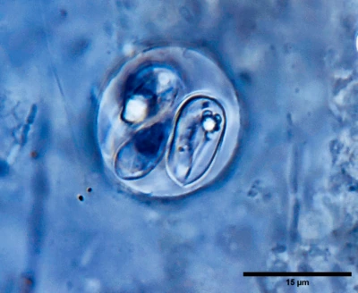
Oocysts are oval, approximately 10–15 µm in size, with a well-defined wall. Inside, four sporocysts can be identified. Their content consists of elongated sporozoites, clearly visible in native preparations.
Under the microscope, oval oocysts with a dense wall are clearly distinguished. Sporocysts occupy most of the internal volume. Notably, the internal organization is well preserved — structures do not disintegrate and retain their shape, which distinguishes them from cellular debris.
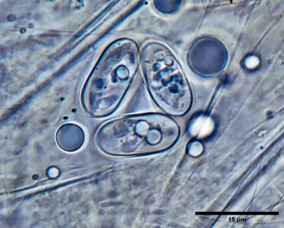
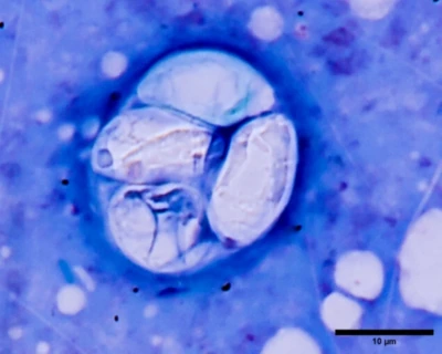
In some preparations, three sporocysts can be observed within a single oocyst. Each contains its own internal structures, including elongated sporozoites. It is important to assess not only the shape of the oocyst, but also its internal architecture — this is what allows differentiation from other inclusions.
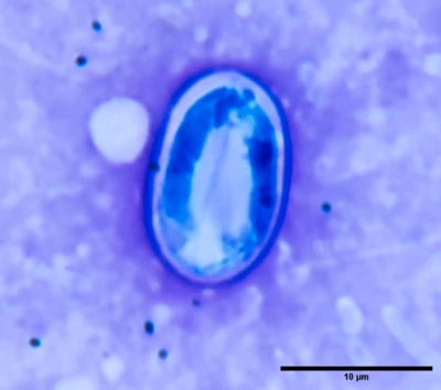
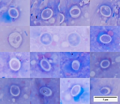
Stained preparations highlight the oocyst wall, although internal structures may become less distinct. In such cases, interpretation should rely on overall shape, size, and repeatability of the observed structures. Uniform staining does not exclude the presence of parasites.
When observing groups of oocysts, the consistency of shape and size becomes evident — one of the key indicators of a parasitic origin. Sporocysts are elongated, and their content is aligned along the longitudinal axis, which is characteristic of Goussia.
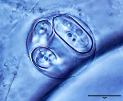
Oocysts with clearly visible sporocysts allow identification of elongated sporozoites. These worm-like structures are among the most reliable diagnostic features and should not be confused with artifacts or degraded tissue.
The key diagnostic feature is the internal organization. In contrast to Eimeria, where sporocysts have a distinct wall and appear more compact, in Goussia sporozoites are seen as structured elongated forms without a clearly separated sporocyst wall.
Parasites localize in the intestinal epithelium and undergo intracellular development. This is associated with epithelial damage, desquamation, and accumulation of mucus in the intestinal lumen. At moderate infection intensity, coccidia may not produce obvious clinical signs. However, with increased parasite load or weakened host condition, digestive disturbances, emaciation, and mortality may occur.
It is important to note that coccidia do not induce granuloma formation. The presence of granulomatous inflammation should prompt investigation of other causes and should not be interpreted as coccidiosis.
Interpretation
The presence of coccidia in the intestine does not always indicate disease. Evaluation of infection intensity and tissue condition is essential.
Single oocysts may be considered incidental findings. A high number of intracellular stages accompanied by epithelial destruction indicates a pathological process.
The presence of sporulating forms within tissue suggests an active life cycle rather than incidental ingestion of oocysts from the environment.
Differential diagnosis
The main challenge is distinguishing Goussia from Eimeria.
Eimeria is characterized by sporocysts with a clearly defined wall. Their internal content typically appears as a compact granular mass.
Goussia, in contrast, lacks a well-defined sporocyst wall, and elongated sporozoites are clearly visible, forming characteristic worm-like structures inside the oocyst.
Cryptosporidium differs by its much smaller size and localization on the epithelial surface, without formation of sporocysts.
Practical relevance
Coccidia are part of the hidden microfauna of aquarium systems. They may persist in a population for long periods without causing overt disease, but under changing conditions can rapidly become clinically significant.
Without microscopy, such processes remain undetected and are often interpreted as “unexplained” disturbances.
Regular monitoring allows early detection of these infections and a more accurate assessment of their significance for fish health.